Test Name
Microscopic Examination for Ehrlichia and Anaplasma (EHRLSM)
CPT Codes
87207
Methodology
Giemsa Prepared Thin Smears
Turnaround Time
7 days
Specimen Requirements
Type:
Whole blood
Volume:
5 mL
Minimum Volume:
1 mL
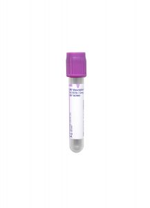
Specimen Container:
Lavender BD Hemogard™ K2EDTA Tube
Transport Temperature:
Ambient
Alternative Specimen
Type:
Whole blood
Volume:
5 mL
Minimum Volume:
1 mL
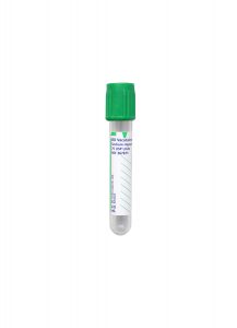
Specimen Container:
Green BD Hemogard™ Sodium Heparin Tubes
Transport Temperature:
Ambient
Stability
Ambient:
24 hours
Refrigerated:
Unacceptable
Frozen:
Unacceptable
Additional Information
Background Information
Ehrlichiosis and anaplasmosis are two closely-related tick-borne diseases caused by different and related small obligate intracellular bacteria. Ehrlichiosis is caused by one of several species of bacteria belonging to the genus Ehrlichia, while anaplasmosis is caused by the bacterium Anaplasma phagocytophilum. Both diseases are concentrated east of the Rocky Mountains, with ehrlichiosis found mainly in mid-Atlantic, southeastern, and south-central states, and anaplasmosis occurring most frequently in the Northeast and upper Midwest areas that are endemic for Lyme disease.
Ehrlichiosis and anaplasmosis are emerging infectious diseases in the United States and other countries and were first recognized as illnesses reportable to the CDC in 1999. According to the National Institute of Allergy and Infectious Disease, 1,009 cases of anaplasmosis, 957 cases of ehrlichiosis, and 132 ehrlichial diseases of undetermined type were reported to the CDC in 2008.
Symptoms often begin one to two weeks following an infected tick bite. Clinical manifestations can be mild or life-threatening, resembling those of Rocky Mountain spotted fever, and are often characterized by sudden high fever, fatigue, chills, muscular aches, and headache.
If left untreated, ehrlichiosis can become a severe, life-threatening disease. Severely-ill patients may have low white blood cell count, anemia, elevated liver enzymes, kidney failure, and respiratory insufficiency; other complications can include seizures and coma. Older patients or those with immune suppression are more likely to require hospitalization. Several deaths have been reported.
Possible complications of anaplasmosis include sepsis and damage to the lungs, heart, kidneys, and nerves. Like ehrlichiosis, the disease is noted for being more severe among individuals with compromised immune systems.
Clinical Indications
A patient with feverish symptoms following tick exposure may suspect ehrlichiosis/anaplasmosis. This test is for individuals who are thought to possibly have ehrlichiosis/ anaplasmosis based on symptoms and clinical presentation.
Morulae may be observed in white blood cells with a peripheral blood smear. Although these intracytoplasmic bodies are not frequently seen, their presence is highly-specific if detected by an experienced microscopist.
Interpretation
A positive result indicates the presence of intracytoplasmic morulae. Determining the type of white blood cell that is infected aids in the differentiation between ehrlichiosis and anaplasmosis.
While the specificity of this assay is high, its sensitivity is limited; a negative result does not exclude the possibility of anaplasmosis or ehrlichiosis.
Methodology
Peripheral blood smear (thin film) review following standard Giemsa staining.
Limitations
This test has limited sensitivity. In rare circumstances, false positives may occur if other intracytoplasmic inclusions are present; however, if the peripheral blood smear is negative, and these diseases are suspected, PCR testing and/or serologic studies are recommended to aid in diagnosis.
References
1. National Institute of Allergy and Infectious Disease. Ehrlichiosis and Anaplasmosis. http://www.niaid.nih.gov/topics/ehrlichiosisanaplasmosis/Pages/Default.aspx. Accessed Feb. 1, 2013.
2. Garcia, Lynne S., ed., et. al. Clinical Microbiology Procedures Handbook, 3rd ed., ASM Press, Washington D.C., 2010, Chapters 2.1.17, 3.4.1.17, 11.7.1-11.7.7.
3. Howard, Barbara J., et. al. Clinical and Pathogenic Microbiology, 2nd ed., Mosby Year Book, St. Louis, 1994, pgs. 859-890.
4. Mahon, Connie R., Textbook of Diagnostic Microbiology, 3rd ed., Saunders Elsevier, St. Louis, Mo., 2007, pgs. 1068-1069.
5. Versalovic, James, ed., et al. Manual of Clinical Microbiology, Vol. 1, 10th edition, ASM Press, Washington D.C., 2011, pgs. 240, 1013-1026.