Posted on

Estimated Glomerular Filtration Rate (eGFR) Updates

Clinical Update

Estimated Glomerular Filtration Rate (eGFR) Updates

The National Kidney Foundation and American Society of Nephrology joint task force recently released recommendations on including a race variable in eGFR reporting (PMID: 34563581).

Based on these recommendations and internal review, the following changes will occur on February 26, 2022:

Adult eGFR Creatinine Calculation

The adult eGFR creatinine equation will be updated from MDRD to the 2021 CKD-EPI creatinine equation in all blood-based creatinine panels (e.g. BMP).

This equation utilizes creatinine, sex, and age, but does not include a race variable.

The new upper reporting limit will increase from 60 to 150 mL/min/1.73m2.

Adult Cystatin C-Based eGFR

eGFR confirmation with a cystatin C-based calculation is recommended near clinical decision-making thresholds.

Two new equations will be available:

2012 eGFR cystatin C (CYSTC)

  • Variables include cystatin C, age, and sex, but not race.

2021 eGFR creatinine-cystatin C (CRECYS)

  • Variables include creatinine, cystatin C, age, and sex, but not race.

Pediatric eGFR Reporting

The task force recommendations focus on the adult population; however, several changes related to EHR transformation will affect the pediatric population:

The Bedside Schwartz creatinine factor will no longer be reported. The Bedside Schwartz creatinine equation will be provided as a comment: Bedside Schwartz equation = 0.413 x [height (cm) / serum creatinine (mg/dL)].

– The 2012 Schwartz cystatin C equation will be utilized for patients 2-17 years old when cystatin C (CYSTC) is ordered.

– The 2021 eGFR creatinine-cystatin C equation (CRECYS) is valid for adults only and will not be available to order for patients less than 18 years of age.

Posted on

September 2021: New Test – Beta(β)-D-Glucan Assay

Clinical Update

New Test: Beta(β)-D-Glucan Assay

In-house Beta(β)-D-Glucan (BDG) testing is now available.

The assay diagnoses certain systemic fungal infections, such as Candidemia, and assists in the prognostication and monitoring response to anti-fungal therapy in hospitalized patients.

This FDA-approved test is a protease zymogen-based colorimetric assay (FUNGITELL® ASSAY) based on a modification of the Limulus Amebocyte Lysate (LAL) pathway in which BDG, where present in the sample, binds Factor G. Activated Factor G turns a proclotting enzyme to clotting enzyme. The latter cleaves an artificial chromogenic substrate (a short peptide) to generate a color. The reaction occurs at 37°C and is kinetically read by a spectrophotometer to generate a standard curve and sample readouts that are subsequently converted to BDG concentrations in pg/ml.

It is important to note that certain fungi, such as the genus Cryptococcus that produces very low levels of BDG, may not result in serum BDG levels sufficiently elevated to be detected by the assay. Infections with fungi of the order Mucorales, such as AbsidiaMucor, and Rhizopus, that are not known to produce BDG can also yield non-detectable serum BDG levels. In addition, the yeast phase of Blastomyces dermatitidis produces little BDG and may not be detected by the assay. Furthermore, positive BDG results have been seen in hemodialysis patients, those who received certain fractionated blood products (such as albumin, IVIg), and those exposed to glucan-containing gauze or surgical sponges.

The sample should be centrifuged within 2 hours of collection to separate the serum from the cells.

Separated samples or serum should be stored refrigerated pending delivery to the testing laboratory.

Heel and fingerprick collections are unacceptable.

Test Name
(1,3) B-D Glucan

Test Code
BDGLUC

CPT Code
87449

Methodology
Protease zymogen-based colorimetric assay (FUNGITELL® ASSAY)

Specimen Requirements
Type: Serum
Volume: 0.5 mL
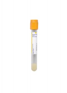
Container: Gold BD Hemogard™ Serum Separation Tubes (SST)™
Transport Temperature: Refrigerated

Stability
Ambient: Unacceptable
Refrigerated: 7 days
Frozen: 1 year

Reference Range
Negative; <60 pg/mL

Reportable Range
31 to 500 pg/mL

Days Performed
Monday, Wednesday, Friday

Posted on

September 2021: Ziad Peerwani, MD Appointed as Medical Director of Cleveland Clinic Laboratories

Clinical Update

Ziad Peerwani, MD Appointed as Medical Director of Cleveland Clinic Laboratories

Ziad Peerwani, MD has been appointed as the new Medical Director for Cleveland Clinic Laboratories, effective September 1, 2021.

Dr. Peerwani was most recently CEO of Union71, based in Abu Dhabi, where he oversaw 17 laboratories, outpatient phlebotomy services, and the performance of more than 15 million lab tests per year.

“We look forward to the experience Dr. Peerwani brings to leading and growing our outreach laboratory services,” says Brian Rubin, MD, PhD, Chair of Cleveland Clinic’s Robert J. Tomsich Pathology & Laboratory Medicine Institute.

Ziad Peerwani, MDD

Ziad Peerwani, MD

About Dr. Peerwani:

In August 2020, Dr. Peerwani was appointed to carve out all laboratory services from SEHA, the government-owned health care system in Abu Dhabi, into an independent company while minimizing the impact on patient care and operations.

After successfully establishing the new business—Union71—Dr. Peerwani was appointed as its first CEO on October 4, 2020. During Union71’s first year, Dr. Peerwani stabilized and grew the laboratory while substantially outperforming Year 1 projections. Eventually, in March 2021, Union71 merged into PureHealth, with Dr. Peerwani supporting another transition.

Before serving as Union71’s CEO, Dr. Peerwani was Chairman of Pathology & Laboratory Medicine at Tawam Hospital, the regional cancer center for SEHA.  He was also the section chief of hematopathology and flow cytometry, providing the majority of clinical sign-out over the years.

Prior to joining Tawam in 2014, Dr. Peerwani practiced general anatomic and clinical pathology at Baylor Scott & White All Saints Medical Center — Fort Worth.

Dr. Peerwani completed his residency and fellowship in Anatomic Pathology, Clinical Pathology, and Hematopathology at Cleveland Clinic.

Posted on

Pathology Insights – Mammary Myofibroblastoma: A Practical Approach with Miglena Komforti, DO

Pathology Insights Video Series

Mammary Myofibroblastoma: A Practical Approach to Pathologic Diagnosis on Core Needle Biopsy

Presented by Miglena Komforti, DO

Myofibroblastoma is an uncommon benign neoplasm of the breast.

Dr. Miglena Komforti, a staff member of Cleveland Clinic’s Breast Pathology Section, discusses the histopathologic findings, ancillary immunohistochemical stains, molecular alterations, and clinical significance of mammary myofibroblastoma.

As part of our educational mission for our clients and communities, Cleveland Clinic Laboratories presents the Pathology Insights video series.
These short videos break down information about interesting pathology cases to better inform doctors, laboratory staff, patients, or anyone interested in the field of pathology. Each episode features important cases, methods, and practices that are personally presented by our staff pathologists.
Posted on

July 2021: Test Menu Optimization & Discontinuations – MGLESE, MYSGRV, PARSYN

Special Communication

Test Menu Optimization & Discontinuations: MGLESE, MYSGRV, & PARSYN

Cleveland Clinic regularly updates its clinical practices to align with the latest testing practices as a part of our Continuous Improvement efforts.

A recent review has identified tests that will be discontinued in favor of more simple, focused, and cost-effective testing options.

Please refer to the test information below or contact Client Services for assistance.

Test Discontinuations

Effective August 3, 2021.

Myasthenia Gravis (MG)/Lambert-Eaton Syndrome (LES) Evaluation, Serum (MGLESE)

Alternative Tests:

Acetylcholine Receptor Binding Antibody Test (ACHRAB)*

ACHRAB Clinical Info:
Anti-acetylcholine receptor binding antibody test is used as an aid in the diagnosis of myasthenia gravis. A negative result cannot exclude myasthenia gravis. Clinical correlation is required.

*Note: Acetylcholine Receptor Modulating Antibody (ACEMOD) and Muscle-Specific Kinase Antibody (MUSK) can be ordered separately if clinically indicated.

P/Q-Type Voltage-Gated Calcium Channel (VGCC) Antibody Test (VOLTCA)

VOLTCA Clinical Info:
Aids in the evaluation of muscle weakness in the context of neuromuscular junction disorder with or without cancer, or the diagnosis of paraneoplastic neurological syndromes.

Myasthenia Gravis Evaluation with Muscle-Specific Kinase (MuSK) Reflex, Serum (MYSGRV)

Alternative Tests:

Acetylcholine Receptor Binding Antibody Test (ACHRAB)*

ACHRAB Clinical Info:
Anti-acetylcholine receptor binding antibody test is used as an aid in the diagnosis of myasthenia gravis. A negative result cannot exclude myasthenia gravis. Clinical correlation is required.

*Note: Acetylcholine Receptor Modulating Antibody (ACEMOD) and Muscle-Specific Kinase Antibody (MUSK) can be ordered separately if clinically indicated.

Paraneoplastic Syndrome Ab Panel with Reflex (PARSYN)

Alternative Tests:

Paraneoplastic Autoantibody Evaluation, Serum (PARNEO)

PARNEO Clinical Info:
This supplemental testing is recommended in cases of chorea, vision loss, cranial neuropathy, and myelopathy.

Posted on

Pathology Insights – Diagnosis of Oncocytic Salivary Gland Tumors with Mobeen Rahman, MD

Pathology Insights Video Series

Diagnosis of Oncocytic Salivary Gland Tumors

Presented by Mobeen Rahman, MD

The diagnosis of oncocytic salivary gland tumors can be challenging, especially on core needle biopsies.

Dr. Mobeen Rahman, staff Head & Neck pathologist, discusses an interesting case that explores this issue.

Follow Dr. Rahman on Twitter: @hnpathology

As part of our educational mission for our clients and communities, Cleveland Clinic Laboratories presents the Pathology Insights video series.
These short videos break down information about interesting pathology cases to better inform doctors, laboratory staff, patients, or anyone interested in the field of pathology. Each episode features important cases, methods, and practices that are personally presented by our staff pathologists.
Posted on

CC-SIGN® Head & Neck Gene Fusion Next-Generation Sequencing Panel

CC-SIGN® Head & Neck Gene Fusion Next-Generation Sequencing Panel

This customized, 30-gene Next-Generation Sequencing (NGS)-based laboratory-developed test is intended for use in the diagnosis and management of benign and malignant tumors of the head and neck.

In particular, salivary gland neoplasms are broadly covered by this panel—which also covers select mesenchymal tumors and other lesions arising in head and neck sites.

The CC-SIGN® Head & Neck Gene Fusion NGS Panel is available as part of a comprehensive, diagnostic consultation or as a stand-alone test. This laboratory-developed test interrogates gene targets associated with known translocations in salivary gland and other solid tumors. Additionally, this panel identifies the corresponding fusion partner which may be helpful in the selection of treatment targets in some cases.

Results are delivered within 14 days of specimen receipt, allowing for a timely, definitive diagnosis in difficult salivary gland tumors and other head and neck lesions in a stand-alone fashion or with the support of our expert pathologists.

Test Overview

Test Name

Head and Neck Next Generation Sequencing (HDNK)

CPT Codes

81445
88381

Turnaround Time

14 days (upon specimen receipt)

Specimen Requirements

Formalin-fixed, paraffin-embedded (FFPE) tissue
• Ten (10) unstained, 4 μM sections of FFPE on charged, unbaked slides
• One (1) H&E stained slide with best tumor area* circled by a pathologist

*minimum of 20% tumor content for best results

Transport Temperature

Room (ambient) temperature

Specimen Shipping Address

Cleveland Clinic Laboratories
2119 E. 93rd Street, L15
Cleveland, OH 44106

Specimens must be sent via UPS, FedEx, or DHL review our Shipping Information for more details.

Clinical Indications

This test is intended for the diagnosis of benign or malignant mesenchymal tumors (sarcomas & their mimics) as well as other solid tumors.

Targeted Gene Regions

Genes interrogated, including relevant transcripts and exons, are listed in alphabetical order.

A

Gene, Transcript, Exons

ALK
NM_004304
2, 4, 6, 10, 16-23, 25, 26

B

Gene, Transcript, Exons

BRAF
NM_004333
Exons 1-5, 7-16, 18

C

Gene, Transcript, Exons

CAMTA1
NM_015215
Exons 3, 8-10

CRTC1
NM_015321
Exons 1-4

E

Gene, Transcript, Exons

ETV6
NM_001987
Exons 1-7

EWSR1
NM_005243
Exons 4-14

F

Gene, Transcript, Exons

FOS
NM_005252
Exon 4

FOSB
NM_006732
Exons 1, 2

FOXO1
NM_002015
Exons 1-3

FUS
NM_004960
Exons 3-11, 13, 14

G

Gene, Transcript, Exons

GLI1
NM_005269
Exons 4-7

H

Gene, Transcript, Exons

HMGA2
NM_003483
Exons 1-5

M

Gene, Transcript, Exons

MAML2
NM_032427
Exons 2, 3

MKL2
NM_014048
Exons 11-13

MYB
NM_001130173
Exons 7-9, 11-16

N

Gene, Transcript, Exons

NCOA1
NM_147223
Exons 12-15

NR4A3
NM_006981
Exon 2

NR4A3
NM_173200
Exons 3, 4

NTRK1*
NM_002529
Exons 2, 4, 6, 8, 10-14

NTRK2*
NM_006180
Exons 5, 7, 9, 11-18

NTRK3*
NM_001007156
Exon 15

NTRK3*
NM_002530
Exons 4, 7, 10, 12-16

NUTM1
NM_175741
Exons 2-4, 6

*A specimen positive for a fusion in one of these genes makes the patient a candidate for larotrectinib treatment.
Standalone NTRK testing is also available via the CC-SIGN® NTRK Gene Fusion NGS Panel.

P

Gene, Transcript, Exons

PAX3
NM_181459
Exons 6-8

PAX7
NM_002584
Exons 6-8

PLAG1
NM_002655
Exons 1-4

PRKD1
NM_002742
Exons 10-13

R

Gene, Transcript, Exons

RET
NM_020630
Exons 2, 4, 6, 11, 15, 16

RET
NM_020975
Exons 8-14

S

Gene, Transcript, Exons

SS18
NM_001007559
Exons 2-6, 8-11

SS18
NM_005637
Exons 2, 3

STAT6
NM_001178078
Exons 1-7, 15-20

T

Gene, Transcript, Exons

TFE3
NM_006521
Exons 2-8

Y

Gene, Transcript, Exons

YAP1
NM_001130145
Exons 1-9

Posted on

Pathology Insights – NUT Carcinoma with Sanjay Mukhopadhyay, MD

Pathology Insights Video Series

NUT Carcinoma

Presented by Sanjay Mukhopadhyay, MD

In this video, Sanjay Mukhopadhyay, MD, Director of Pulmonary Pathology at Cleveland Clinic, highlights a rare, aggressive, malignant tumor and draws attention to a distinctive immunohistochemical profile that can serve as a tip-off to the correct diagnosis.

As part of our educational mission for our clients and communities, Cleveland Clinic Laboratories presents the Pathology Insights video series.
These short videos break down information about interesting pathology cases to better inform doctors, laboratory staff, patients, or anyone interested in the field of pathology. Each episode features important cases, methods, and practices that are personally presented by our staff pathologists.
Posted on

Pathology Insights – Diagnosis of CD30+ T-cell Lymphoproliferative Disorders with Genevieve Crane, MD, PhD

Pathology Insights Video Series

Diagnosis of CD30+ T-cell Lymphoproliferative Disorders

Presented by Genevieve Crane, MD, PhD

The diagnosis of CD30+ T-cell lymphoproliferative disorders, including ALK+ and ALK-negative forms of anaplastic large cell lymphoma, can present challenges. These entities show overlapping morphologic and immunohistochemical features but can vary widely in terms of clinical aggression.

Dr. Genevieve Crane, Staff Hematopathologist at Cleveland Clinic, discusses clinical presentation, morphologic features, and emerging prognostic markers that may aid in diagnosis and clinical management.

As part of our educational mission for our clients and communities, Cleveland Clinic Laboratories presents the Pathology Insights video series.
These short videos break down information about interesting pathology cases to better inform doctors, laboratory staff, patients, or anyone interested in the field of pathology. Each episode features important cases, methods, and practices that are personally presented by our staff pathologists.
Posted on

Pathology Insights – Two Diagnoses in One Bone Marrow with Megan Nakashima, MD

Pathology Insights Video Series

Two Diagnoses in One Bone Marrow

Presented by Megan Nakashima, MD

Analysis of a bone marrow requires both careful morphologic examination as well as appropriate ancillary studies.

Dr. Megan Nakashima, Staff Hematopathologist at Cleveland Clinic, discusses a complicated bone marrow case involving a patient with thrombocytopenia, monocytosis, and diarrhea. After requiring the incorporation of molecular and immunophenotypic information, two diagnoses are reached.

In addition to this case, Dr. Nakashima reviews relevant changes in the WHO 2016 classification of myeloid and associated neoplasms.

As part of our educational mission for our clients and communities, Cleveland Clinic Laboratories presents the Pathology Insights video series.
These short videos break down information about interesting pathology cases to better inform doctors, laboratory staff, patients, or anyone interested in the field of pathology. Each episode features important cases, methods, and practices that are personally presented by our staff pathologists.