Test Name
CPT Code
81445
Methodology
Next-Generation Sequencing (NGS)
Turnaround Time
8 days
Specimen Requirements
Type:
FFPE Tissue
Volume:
– 15 charged, unbaked, and unstained slides sectioned at 7 μm.
– One pre and one post H&E slide with tumor area circled, and percent tumor indicated.
Transport Temperature:
Ambient
Type:
Cytology
Volume:
– 15 charged, unbaked and unstained slides sectioned at 7 μm.
– One H&E slide with tumor percent indicated.
Transport Temperature:
Ambient
Type:
Bone Marrow Aspirate
Volume:
2 mL
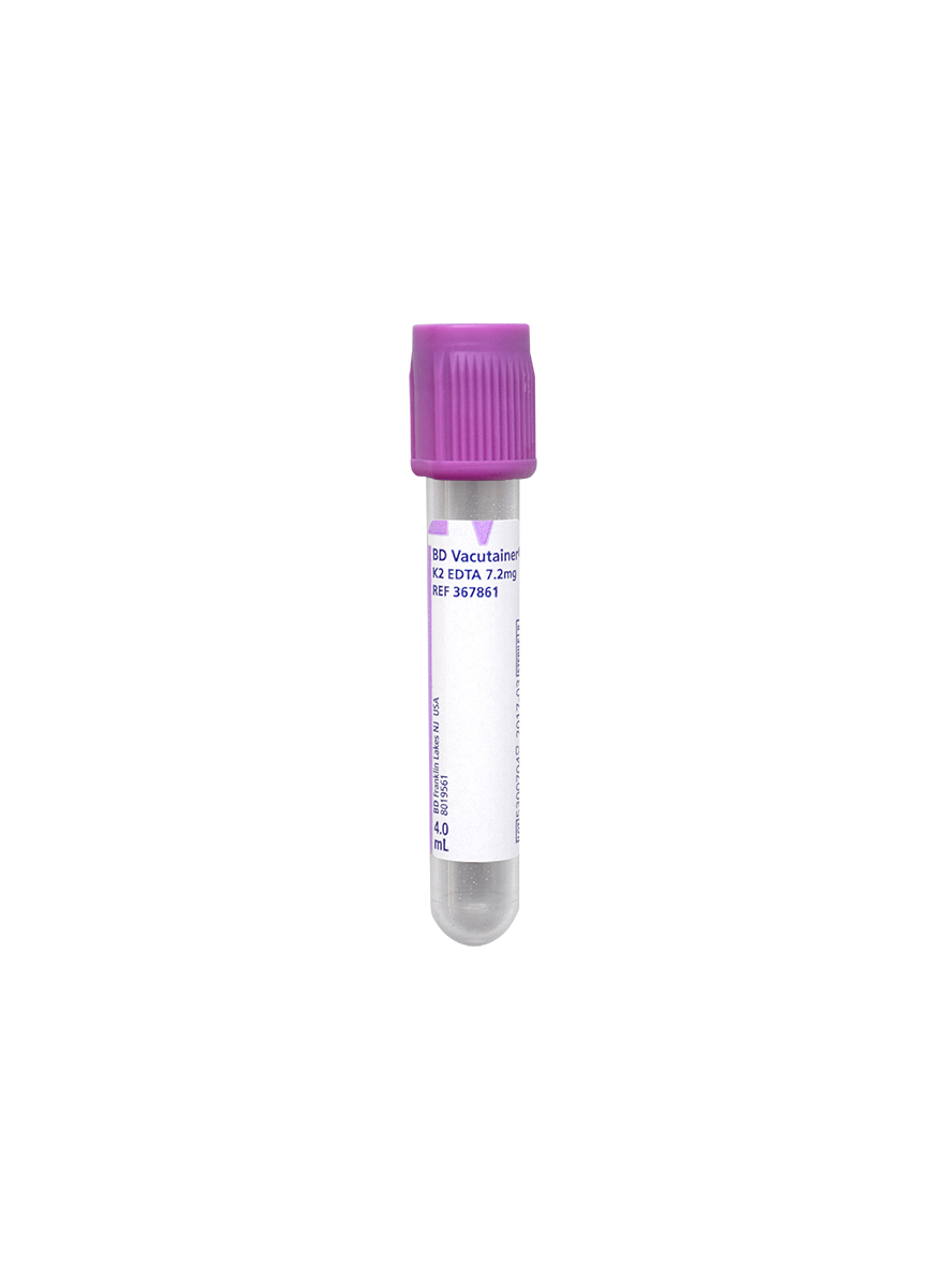
Type:
Peripheral blood
Volume:
4 mL
Tube/Container:
Lavender BD Hemogard™ K2EDTA Tubes
Transport Temperature:
Ambient
Stability
FFPE Tissue
Ambient:
Indefinitely
Refrigerated:
Indefinitely
Frozen:
Unacceptable
Cytology Alcohol or Formalin-Fixed Cell Block
Ambient:
Indefinitely
Refrigerated:
Indefinitely
Frozen:
Unacceptable
Bone Marrow or Peripheral Blood
Ambient:
48 hours
Refrigerated:
3 days
Frozen:
Unacceptable
Additional Information
Overview
Recurrent somatic alterations are found in a variety of solid tumors. Identifying such alterations provides pathologists and clinicians with valuable data that may assist in the diagnosis, classification, prognostic evaluation, and therapeutic management of these malignancies.
The Targeted Oncology Panel (TOP) is a custom 59-gene Next-Generation Sequencing (NGS) panel developed for the identification of single nucleotide variants (SNVs), small insertions and deletions (indels), and copy number gains (CNVs) from DNA specimens, and fusion transcripts and aberrant transcripts from RNA specimens. The workflow allows for the concurrent testing of DNA and RNA to analyze over 1,000 biomarkers.
This panel is optimized to evaluate scant specimens, such as cytology specimens and limited biopsies; as such, TOP may be used for advanced tumor profiling and clinical trial evaluation when material is too limited to send for comprehensive sequencing. For example, TOP can detect resistance mutations in EGFR, KRAS, NRAS, and PDGFRA, as well as evolving biomarkers in AKT1, BRAF non-V600, CDK4, ERBB2, HRAS, MAP2K1/2, and MTOR, amongst others.
Besides providing useful information for cancer evaluation and diagnosis, this test can identify pan-cancer therapeutic markers, including:
TOP Biomarker
FDA-Approved Treatment Examples*
ALK fusions
Alectinib, Brigatinib, Ceritinib, Crizotinib for NSCLC
ALK oncogenic mutations
Lorlatinib for NSCLC
BRAF V600E
Vemurafenib, Dabrafenib and others for Melanoma
Dabrafenib + Trametinib for all solid tumors
EGFR exon 19 in-frame deletions
Gefitinib, Osimertinib for NSCLC
EGFR exon 20 in-frame insertions
Amivantamab, Mobocertinib for NSCLC
ERBB2 (HER2) amplification
Multiple therapy options for Breast Cancer
Tucatinib + Trastuzumab for CRC
Trastuzumab options for Esophagogastric Cancer
FGFR2 fusions
Erdafitinib for Bladder Cancer
Futibatinib, Pemigatinib for Cholangiocarcinoma
FGFR3 fusions or activating mutations
Erdafitinib for Bladder Cancer
IDH1 R132
Ivosidenib for Cholangiocarcinoma
KIT oncogenic mutations
Imatinib, Regorafenib, Ripretinib, Sunitinib for GIST
KRAS G12C
Adagrasib, Sotorasib for NSCLC
MET exon 14 skipping alterations or amplification
Capmatinib, Tepotinib for NSCLC
NTRK1/2/3 fusions
Entrectinib, Larotrectinib for all solid tumors
PDGFRA exon 18 alterations
Avapritinib for GIST
PIK3CA oncogenic mutations
Alpelisib + Fulvestrant in Breast Cancer
RET fusions
Selpercatinib for all tumors
RET mutations
Pralsetinib, Selpercatinib for Medullary Thyroid Cancer
ROS1 fusions
Crizotinib, Entrectinib for NSCLC
Clinical Indications
Hotspots, copy number variants, and selected fusions evaluated by this assay can aid in the diagnostic and therapeutic assessment of a variety of tumor types, including non-small cell lung cancer, melanoma, colorectal cancer, prostate cancer, breast cancer, glioblastoma, thyroid cancer, and others.
This panel can also provide focused tumor profiling for patients with locally advanced/metastatic disease, who are candidates for anti-cancer therapy, to identify uncommon but targetable alterations.
Interpretation
Variants are classified according to established guidelines, and an interpretation is provided. Reported variants include those of strong or potential clinical significance and variants of unclear clinical significance.
Common population variants are not included in the report.
Methodology & Limitations
Extracted nucleic acid from the specimen, both DNA and RNA, were subjected to separate targeted amplification reactions using AmpliSeq custom primers designed by Thermo Fisher Scientific (Thermo Fisher Scientific, Waltham, MA). Hotspots and selected fusions in gene regions listed below were sequenced using Illumina (San Diego, CA) 2×150 paired-end cycle chemistry. A customized bioinformatics analytical platform was used for read alignment (Genome Build GRCh37/hg19), variant identification, and annotation. Single nucleotide variants (SNVs), insertion, deletion (indels), and copy number gain variants are detected by DNA sequencing. Select fusions and aberrant transcripts (EGFR vIII and MET exon 14 skipping transcripts) are detected by RNA sequencing. Variants are classified according to established guidelines. Reported results include variants of strong or potential clinical significance and variants of unclear clinical significance. Benign population polymorphisms are not included in the report.
Based on validation, the DNA testing delivered an average of >500x coverage, and >99% of targeted regions showed over 100x coverage. A minimum coverage depth of 100 reads is required across the entire region of interest; a list of low-coverage areas is included in the report as applicable. The test demonstrated 100% sensitivity and 100% specificity in identifying SNVs, indels, and copy number gains. The lower limit of detection of this assay is approximately 5% variant allele fraction (VAF) for SNV/indels and 6 copies or greater for copy number gains. Variants below these thresholds may be reported at the discretion of the molecular pathology professional staff if the technical quality of the sequencing is sufficient at that location and the call is unequivocal.
Based on validation, the RNA fusion testing averaged >150,000 total reads. The test demonstrated 93%sensitivity and 100% specificity in gene fusion identification compared to NGS sequencing and 69% sensitivity and 100% specificity compared to fluorescence in situ hybridization (FISH) of fusion drivers (unknown fusion partner). Overall sensitivity is 78%, and accuracy is 99%. The lower limit of detection is approximately 1% of total sequencing reads.
Sequence changes outside the analyzed alterations hotspots, including intronic and noncoding regions, will not be identified by this test. Insertions and deletions larger than 20 and 40 bp, respectively, may not be identified by this test. Negative results from specimens for which the percentage of tumor cells is 10% or less should be interpreted with caution. Although variant allele fraction is provided as a percentage, this is not a quantitative test. RNA fusions involving alternative partners or breakpoints outside of the targeted regions cannot be detected by this test. This test does not distinguish between somatic and inherited variants. Tumor heterogeneity, tumor burden, specimen degradation or other limitations of the technology may affect the sensitivity and limit of detection, either broadly across the regions of interest or for specific regions, and may lead to false negative results. This test is not intended to be used for circulating tumor evaluation.
Genes & Hotspot Regions
Genes & Hotspot Regions Covered for SNV/Indels:
| wdt_ID | Gene | Transcript | Exon(s)* |
|---|---|---|---|
| 1 | AKT1 | NM_005163 | 3 |
| 2 | ALK | NM_004304 | 21-25 |
| 3 | AR | NM_000044 | 6, 8 |
| 4 | BRAF | NM_004333 | 11, 15 |
| 5 | CDK4 | NM_000075 | 2 |
| 6 | CTNNB1 | NM_001904 | 3, 7-8 |
| 7 | DDR2 | NM_006182 | 5 |
| 8 | EGFR | NM_005228 | 3 ,7, 12, 15, 18-21 |
| 9 | ERBB2 | NM_004448 | 8, 17-22 |
| 10 | ERBB3 | NM_001982 | 2-3, 6, 8-9 |
| 11 | ERBB4 | NM_005235 | 18 |
| 12 | ESR1 | NM_000125 | 8 |
| 13 | FGFR1 | NM_023110 | 12-14, 16-17 |
| 14 | FGFR2 | NM_000141 | 7-9, 12, 14 |
| 15 | FGFR3 | NM_000142 | 7, 9, 14, 16 |
| 16 | GNA11 | NM_002067 | 4, 5 |
| 17 | GNAQ | NM_002072 | 4, 5 |
| 18 | GNAS | NM_080425 | 8 |
| 19 | H3-3A | NM_002107 | 2 |
| 20 | H3-3B | NM_005324 | 2 |
| 21 | HRAS | NM_005343 | 2, 3 |
| 22 | IDH1 | NM_005896 | 4 |
| 23 | IDH2 | NM_002168 | 4 |
| 24 | JAK1 | NM_002227 | 14-16 |
| 25 | JAK2 | NM_004972 | 14 |
| 26 | JAK3 | NM_000215 | 11-12, 15 |
| 27 | KIT | NM_000222 | 8-11, 13, 17 |
| 28 | KRAS | NM_033360 | 2-4 |
| 29 | MAP2K1 | NM_002755 | 2-3, 6 |
| 30 | MAP2K2 | NM_030662 | 2 |
| 31 | MET | NM_000245 | 14, 16, 19 |
| 32 | MTOR | NM_004958 | 30, 39-40, 43, 47, 53 |
| 33 | NRAS | NM_002524 | 2-4 |
| 34 | PDGFRA | NM_006206 | 12, 14, 18 |
| 35 | PIK3CA | NM_006218 | 2, 5-6, 8, 10, 14, 19, 21 |
| 36 | RAF1 | NM_002880 | 7, 12 |
| 37 | RET | NM_020975 | 11, 13, 15-16 |
| 38 | ROS1 | NM_002944 | 36, 38 |
| 39 | SMO | NM_005631 | 4, 6, 8-9 |
| 40 | TERT | NM_198253 | Promoter |
| 41 | TP53 | NM_000546 | 5, 7, 8 |
Fusions Detected in RNA
Genes reported for copy number gains: ALK, AR, BRAF, CCND1, CDK4, CDK6, EGFR, ERBB2, FGFR1, FGFR2, FGFR3, FGFR4, KIT, KRAS, MET, MYC, MYCN, PDGFRA, PIK3CA
TOP RNA Fusions
| wdt_ID | Gene | Detected Fusions |
|---|---|---|
| 1 | ABL1 | EML1::ABL1 |
| 2 | AKT3 | MAGI3::AKT3 |
| 3 | ALK | A2M::ALK, ACTG2::ALK, ALK::PTPN3, ATIC::ALK, C2orf44::ALK, CARS::ALK, CLIP4::ALK, CLTC::ALK, DCTN1::ALK, EML4::ALK, GTF2IRD1::ALK, HIP1::ALK, KIF5B::ALK, KLC1::ALK, MEMO1::ALK, NCOA1::ALK, PRKAR1A::ALK, RANBP2::ALK, SEC31L1_SEC31A::ALK, SMEK2::ALK, STRN:: |
| 4 | AXL | AXL::MBIP |
| 5 | BRAF | AGTRAP::BRAF, AKAP9::BRAF, CDC27::BRAF, FAM131B::BRAF, FCHSD1::BRAF, KIAA1549::BRAF, PAPSS1::BRAF, SLC45A3::BRAF, SND1::BRAF, TAX1BP1::BRAF, TRIM24::BRAF |
| 6 | EGFR | EGFR vIII transcript |
| 7 | ERBB2 | WIPF2::ERBB2 |
| 8 | ERG | SLC45A3::ERG, TMPRSS2::ERG |
| 9 | FGFR1 | BAG4::FGFR1, ERLIN2::FGFR1, FGFR1::TACC1 |
| 10 | FGFR2 | FGFR2::AFF3, FGFR2::BICC1, FGFR2::CASP7, FGFR2::CIT, FGFR2::KIAA1967_CCAR2, FGFR2::MGEA5, FGFR2::OFD1, FGFR2::TACC1, SLC45A3::FGFR2 |
| 11 | FGFR3 | FGFR3::AES, FGFR3::BAIAP2L1, FGFR3::ELAVL3, FGFR3::TACC3 |
| 12 | MET | MET Exon 14 Skipping, BAIAP2L1::MET, C8orf34::MET, CAPZA2::MET, OXR1::MET, TFG::MET, TPR::MET, PTPRZ1::MET |
| 13 | NTRK1 | BCAN::NTRK1, CD74::NTRK1, CEL::NTRK1, IRF2BP2::NTRK1, LMNA::NTRK1, MPRIP::NTRK1, NFASC::NTRK1, NTRK1::DYNC2H1, RNF213::NTRK1, SQSTM1::NTRK1, SSBP2::NTRK1, TFG::NTRK1, TPM3::NTRK1, TPR::NTRK1 |
| 14 | NTRK2 | AFAP1::NTRK2, AGBL4::NTRK2, NACC2::NTRK2, QKI::NTRK2, SQSTM1::NTRK2, TRIM24::NTRK2, VCL::NTRK2 |
| 15 | NTRK3 | BTBD1::NTRK3, COX5A::NTRK3, ETV6::NTRK3 |
| 16 | PDGFRA | SCAF11::PDGFRA |
| 17 | PPARG | PAX8::PPARG |
| 18 | RAF1 | B4GALT1::RAF1, ESRP1::RAF1 |
| 19 | RELA | C11orf95::RELA |
| 20 | RET | ACBD5::RET, AFAP1::RET, AKAP13::RET, CCDC6::RET, CUX1::RET, ERC1::RET, FKBP15::RET, GOLGA5::RET, HOOK3::RET, KIAA1468::RET, KIF5B::RET, KTN1::RET, NCOA4::RET, PCM1::RET, PRKAR1A::RET, RUFY2::RET, SPECC1L::RET, TBL1XR1::RET, TRIM24::RET, TRIM27::RET, TRIM3 |
| 21 | ROS1 | CCDC6::ROS1, CD74::ROS1, CEP85L::ROS1, CLIP1::ROS1, CLTC::ROS1, ERC1::ROS1, EZR::ROS1, GOPC::ROS1, HLA_A::ROS1, KDELR2::ROS1, KIAA1598::ROS1, LRIG3::ROS1, MSN::ROS1, MYO5A::ROS1, PPFIBP1::ROS1, PWWP2A::ROS1, SDC4::ROS1, SLC34A2::ROS1, TFG::ROS1, TPM3::ROS |
| 22 | TMPRSS2 | TMPRSS2::ERG, TMPRSS2::ETV1, TMPRSS2::ETV4, TMPRSS2::ETV5 |
References
1. Li MM, et al. Standards and Guidelines for the Interpretation and Reporting of Sequence Variants in Cancer: A Joint Consensus Recommendation of the Association for Molecular Pathology, American Society of Clinical Oncology, and College of American Pathologists. J Mol Diagn. 2017 Jan;19(1):4-23. doi: 10.1016/j. jmoldx.2016.10.002. PMID: 27993330; PMCID: PMC5707196.
2. Kumar-Sinha C, Tomlins SA, Chinnaiyan AM. Recurrent gene fusions in prostate cancer. Nat Rev Cancer. 2008 Jul;8(7):497-511. doi: 10.1038/nrc2402. Epub 2008 Jun 19. PMID: 18563191; PMCID: PMC2711688.
3. Baca SC, Garraway LA. The genomic landscape of prostate cancer. Front Endocrinol (Lausanne). 2012 May 16;3:69. doi: 10.3389/fendo.2012.00069. PMID: 22649426; PMCID: PMC3355898.
4. Bennett C, et al. Breast Cancer Genomics: Primary and Most Common Metastases. Cancers (Basel). 2022 Jun 21;14(13):3046. doi: 10.3390/cancers14133046. PMID: 35804819; PMCID: PMC9265113.
5. Natrajan R, Tutt ANJ, Lord CJ. Driver Oncogenes but Not as We Know Them: Targetable Fusion Genes in Breast Cancer. Cancer Discov. 2018 Mar;8(3):272-275. doi: 10.1158/2159-8290.CD-18-0091. PMID: 29500329; PMCID: PMC6372062.
6. Singh A, Ham J, Po JW, Niles N, Roberts T, Lee CS. The Genomic Landscape of Thyroid Cancer Tumourigenesis and Implications for Immunotherapy. Cells. 2021 May 1;10(5):1082. doi: 10.3390/cells10051082. PMID: 34062862; PMCID: PMC8147376.
7. Sepulveda AR, et al. Molecular Biomarkers for the Evaluation of Colorectal Cancer: Guideline From the American Society for Clinical Pathology, College of American Pathologists, Association for Molecular Pathology, and the American Society of Clinical Oncology. J Clin Oncol. 2017 May 1;35(13):1453-1486. doi: 10.1200/JCO.2016.71.9807. Epub 2017 Feb 6. PMID: 28165299.
8. Lindeman NI, et al. Updated Molecular Testing Guideline for the Selection of Lung Cancer Patients for Treatment With Targeted Tyrosine Kinase Inhibitors: Guideline From the College of American Pathologists, the International Association for the Study of Lung Cancer, and the Association for Molecular Pathology. Arch Pathol Lab Med. 2018 Mar;142(3):321-346. doi: 10.5858/arpa.2017-0388-CP. Epub 2018 Jan 22. PMID: 29355391.
9. Cancer Genome Atlas Research Network. Comprehensive genomic characterization defines human glioblastoma genes and core pathways. Nature. 2008 Oct 23;455(7216):1061-8. doi: 10.1038/nature07385. Epub 2008 Sep 4. Erratum in: Nature. 2013 Feb 28;494(7438):506. PMID: 18772890; PMCID: PMC2671642.
10. Lombardi MY, Assem M. Glioblastoma Genomics: A Very Complicated Story. In: De Vleeschouwer S, editor. Glioblastoma [Internet]. Brisbane (AU): Codon Publications; 2017 Sep 27. Chapter 1. PMID: 29251861.
11 .Guhan S, Klebanov N, Tsao H. Melanoma genomics: a state-of-the-art review of practical clinical applications. Br J Dermatol. 2021 Aug;185(2):272-281. doi: 10.1111/bjd.20421. Epub 2021 Jun 6. PMID: 34096042.
12. Tornillo L, Terracciano LM. An update on molecular genetics of gastrointestinal stromal tumours. J Clin Pathol. 2006 Jun;59(6):557-63. doi: 10.1136/jcp.2005.031112. PMID: 16731599; PMCID: PMC1860404.
13. Malone ER, Oliva M, Sabatini PJB, Stockley TL, Siu LL. Molecular profiling for precision cancer therapies. Genome Med. 2020 Jan 14;12(1):8. doi: 10.1186/s13073-019-0703-1. PMID: 31937368; PMCID: PMC6961404.
14. Chakravarty D et al. Somatic Genomic Testing in Patients With Metastatic or Advanced Cancer: ASCO Provisional Clinical Opinion. J Clin Oncol. 2022 Apr 10;40(11):1231-1258. doi: 10.1200/JCO.21.02767. Epub 2022 Feb 17. Erratum in: J Clin Oncol. 2022 Jun 20;40(18):2068. PMID: 35175857.