Test Name
Colagen Binding Assay (CBA)
CPT Codes
83520
Methodology
Enzyme-Linked Immunosorbent Assay (ELISA)
Turnaround Time
1 – 3 days
Specimen Requirements
Volume:
2 mL
Specimen Type:
Plasma
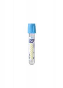
Collection Container:
Light Blue Sodium Citrate Coagulation Tube
Transport Temperature:
Frozen
Specimen Collection & Transport
Collection of blood by routine venipuncture in a 3.5 mL light blue top tube containing a 9:1 ratio of blood to 3.2% trisodium citrate anticoagulant.
Pediatric volume of 2.5 mL with an appropriate ratio of anticoagulant is acceptable.
Reference Range
CBA:
41-161%
Ratio of CBA/VWF:
Ag >=0.73
Additional Information
Background Information
Von Willebrand disease (VWD) is the most common inherited bleeding disorder with a prevalence of approximately 1% in the general population. It also can occur as an acquired bleeding disorder. VWD is a clinically heterogeneous disorder with several subtypes due to deficiency and/or dysfunction of von Willebrand factor (VWF). VWF is a multimeric adhesive glycoprotein that plays a major role in primary hemostasis and coagulation. VWF mediates adhesion of platelets to injured subendothelium and to the platelet surface receptor GPIb and serves as the specific carrier protein for coagulation factor VIII (fVIII) in plasma preventing proteolytic degradation. The revised classification of VWD identified two major categories: quantitative and qualitative defects.
The quantitative VWF defects include type 1 (partial deficiency of VWF) and type 3 (complete absence of VWF) in plasma and/or platelets. Type 2 is a qualitative VWF defect that is further classified as four subtypes by different pathophysiologic mechanisms.
Accurate laboratory diagnosis and classification of VWD using both quantitative (antigenic) and qualitative (functional) assays based on the VWD diagnostic algorithm are crucial because the presenting biological activity of VWF determines both the hemorrhagic risk and subsequent clinical management.

Abbreviations:
Ag: Antigen
CBA: Collagen-binding activity
fVIIl:C: Factor VIII: Coagulant
MW: Molecular weight
NL: Normal
PFA-100: Platelet function screen
PT: Prothrombin time
PTT: Partial thromboplastin time
RiCof: Ristocetin cofactor
RIPA: Ristocetin-induced platelet aggregation
VWF: von Willebrand factor
Clinical Indications
The functional activity of VWF traditionally has been assessed using the ristocetin cofactor activity (RiCof) assay, which measures the VWF-mediated agglutination of platelets in the presence of ristocetin. However, the usefulness of this assay has limitations due to poor reproducibility and lack of calibration. The collagen-binding activity (CBA) assay has been proposed as a supplemental test for VWF activity.
The CBA assay is based on the ability of multimeric forms of VWF to bind collagen, and its greatest strength lies in the ability to selectively detect primarily high molecular weight (HMW) forms of VWF, which are known to be most functional and adhesive.
The CBA assay is a useful adjunctive to the RiCof assay for the diagnosis of VWD and to differentiate VWD with deficiency of HMW multimer forms in type 2A and type 2B from type 1. It also can differentiate very low levels of VWF in severe type 1 from a complete absence of VWF in type 3 and has been reported as a better marker for therapeutic efficacy of treatment with DDAVP® (desmopressin) and fVIII concentrate.
Interpretation
CBA results are reported as % of the reference value for CBA. CBA to VWF:Ag ratio is calculated to provide a ratio of VWF activity to protein amount.
1. Type 1 VWD patients have concordantly decreased CBA and VWF:Ag levels.
2. Type 3 VWD patients have markedly decreased or nearly absent CBA and VWF:Ag levels.
3. Type 2A VWD and type 2B VWD patients have discordantly decreased CBA and VWF:Ag levels with markedly decreased CBA level, normal, or decreased VWF:Ag level and loss of HMW multimers.
4. Type 2M VWD patients have a discordantly decreased CBA level with a normal or decreased VWF:Ag level but without the loss of HMW multimers.
5. Type 2N VWD patients have normal CBA and VWF:Ag with discordantly decreased FVIII coagulant activity.
6. CBA values are known to be lower in O blood groups compared with non-O blood groups. However, as VWF:Ag levels show similar blood group dependence, the ratio of CBA/VWF:Ag is not affected.
Methodology
The CBA assay is an enzyme immunoassay (REAADS® Collagen Binding Assay ELISA kit, Corgenix, Inc., Broomfield, Colo.) that quantitates the binding of VWF to a collagen-coated microwell plate. After binding peroxidase-conjugated anti-VWF antibodies to VWF multimers, the resulting color intensity is determined photometrically, which is proportional to HMW forms of VWF present in the plasma. In situ evaluation for precision and accuracy of the CBA assay shows low coefficient of variation (6.3-11.1%) with a lower limit of detection 0.2% (linearity 1-530%).
Suggested Reading
1. Favaloro EJ. Von Willebrand factor collagen-binding (activity) assay in the diagnosis of von Willebrand disease: a 15-year journey. Seminar Thromb Hemost. 2002;28:191-202.
2. Nichols WL et al. The diagnosis, evaluation, and management of von Willebrand disease. U.S. Department of Health and Human Services. NIH Publication No. 08-5832, December 2007.
3. Castaman G, Federici AB, Rodeghiero F, Mannucci PM. Von Willebrand disease in the year 2003: toward the complete identification of gene defects for correct diagnosis and treatment. Haematologica. 2003;Jan;88(1):94-108.
4. Kalla A, Talpsep T. The von Willebrand factor collagen binding activity assay: clinical application. An Hematol. 2001;80:466-471.
5. Favaloro EJ. Toward a new paradigm for the identification and functional characterization of Von Willebrand disease. Seminar Thromb Hemost. 2009;35:60-75.