Specimen Collection & Transport
Medical & Transplant Kidney Biopsy Consultation Kit Instructions
Renal biopsy is a useful tool in evaluating patients with a wide variety of kidney diseases.
A comprehensive panel of histochemical stains, immunofluorescence studies, and electron microscopy are all important in the full evaluation of medical and transplant kidney biopsies.
Renal biopsies are most impactful for the patient and treating physician when pertinent clinical information, adequate tissue, and properly shipped specimens are provided to the renal pathologist reviewing the case.
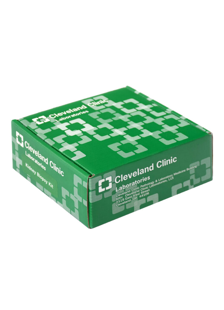
Cleveland Clinic’s Medical & Transplant Kidney Biopsy Consultation Kit provides the necessary materials to ensure specimen integrity during shipment to our laboratories.
Specimen Collection & Transport
Overview
Renal biopsy is a useful tool in evaluating patients with a wide variety of kidney diseases. Patients who have received a kidney transplant undergo biopsies to monitor for transplant rejection. Patients who have not received a transplant but have hypertension, diabetes, autoimmune diseases, infections, or other underlying illnesses can also develop kidney disease.
Determining the cause of kidney disease often helps to guide clinical treatment. Renal biopsies for this purpose are sometimes referred to as “random” kidney biopsies or “medical” kidney biopsies to differentiate from those that may be performed in the evaluation of a renal mass. These biopsies can be requested by a nephrologist or rheumatologist and performed by either an interventional radiologist or nephrologist.
These renal biopsy specimens require highly specialized processing techniques and evaluation by a renal pathologist. A comprehensive panel of histochemical stains, immunofluorescence studies, and often electron microscopy are all important in the full evaluation of medical and transplant kidney biopsies. Ultimately, it is the job of the renal pathologist to combine the diagnostic information provided by these special processing techniques and interpret any findings in the clinical context in which the biopsy was performed.
Renal biopsies are most impactful for the patient and treating physician when pertinent clinical information, adequate tissue, and properly shipped specimens are provided to the renal pathologist reviewing the case.
Requisition
Prior to specimen collection, the treating physician should obtain and complete a Medical & Transplant Kidney Biopsy Requisition.
When filling out the referring physician contact section, please provide contact information for the physician using the biopsy results to guide clinical decision-making.
Complicated cases often require a conversation between the renal pathologist and the treating physician to maximize the diagnostic information gained from the biopsy material.
Specimen Requirements
Obtaining an adequate amount of renal cortex is critical for accurate diagnosis. Separate portions of renal cortex are needed for light microscopy, immunofluorescence, and electron microscopy.
Two cores of tissue, 1-2 cm in length, composed predominantly of renal cortex, will generally yield adequate glomeruli.
In typical cases:
At least 10 glomeruli are needed for light microscopy.
3–5 glomeruli are needed for immunofluorescence.
3–5 glomeruli are needed for electron microscopy.
The physician performing the biopsy should place the specimen on Telfa gauze moistened with saline. If possible, confirm that an adequate amount of glomeruli have been sampled in case additional cores of tissue are needed to be obtained during the procedure.
Deliver the specimen promptly to the local pathology laboratory, where it can be appropriately divided and placed into the appropriate fixatives and transport solutions.
The Medical & Transplant Kidney Biopsy Requisition should accompany the specimen.
Laboratory Handling

10% Neutral Buffered Formalin
Used for light microscopy specimens.
Michel’s (or Zeus) Solution
Used for immunofluorescence specimens.
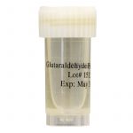
2-4% Glutaraldehyde
Used for electron microscopy specimens (if applicable).
Dividing the Cortex and Medulla
If using a microscope or hand lens to differentiate between cortex and medulla:
1. Place at least half of the cortical tissue in formalin for light microscopy (ideally more than 10 glomeruli).
2. Evenly divide the remaining cortex, ideally 6-10 glomeruli, in Michel’s (or Zeus) fixative for immunofluorescence and glutaraldehyde for electron microscopy.
Alternate Specimen Dividing Technique
If not using a technique for distinguishing cortex and medulla, please use the following empiric technique to divide the specimen:
1. Cut a 1 mm portion of tissue from both ends of each tissue core and place it in glutaraldehyde.
2. Place 1/3 of what remains of each tissue core in Michel’s solution.
3. Place the remaining tissue (which should be at least half of the overall specimen) in formalin.
Dividing Limited/Suboptimal Specimens
Sometimes, there may not be adequate tissue to divide into three parts, and judgment must be exercised as to which diagnostic studies are most useful.
Light microscopy is of the highest priority in almost all cases, and the best tissue should be placed in formalin.
After light microscopy, immunofluorescence is typically most helpful; therefore, if possible, a small portion of the cortex should be placed in Michel’s or Zeus solution.
Renal pathologists can sometimes perform electron microscopy on tissue after being used for light microscopy and immunofluorescence as a salvage technique; thus, electron microscopy should usually be the lowest priority when dividing limited tissue. Note: The main exception to this would be in the setting of pediatric nephrotic syndrome, where electron microscopy is typically more critical than immunofluorescence.
Electron microscopy is typically not performed for transplant biopsies unless there is substantial proteinuria. However, if tissue is limited, medulla can be used for immunofluorescence to evaluate for antibody-mediated rejection. Therefore, when tissue is limited in the transplant setting, please prioritize light microscopy over all other modalities, and put a portion of medulla in Michel’s or Zeus solution for immunofluorescence.
Shipping Preparation
1. After dividing the specimens into the appropriate containers, label each container with two of three patient identifiers:
- Patient name
- Date of birth
- Social security number
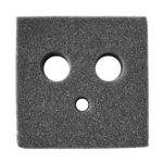
2. Place all specimens into the appropriate openings in the grey foam insert.
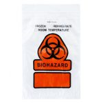
3. Place the foam insert into the provided sealable biohazard bag.
4. Secure the bagged foam insert with the specimens into the green shipping box.
5. Place the completed Medical & Transplant Kidney Biopsy requisition in the Laboratory Pak.
6. Seal the specimens into the Laboratory Pak.

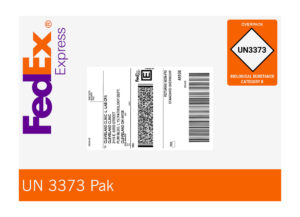
7. Affix the provided shipping label to the Laboratory Pak.
- Ship the specimen Next-Day Air to:
Cleveland Clinic Laboratories
L15 – Pathology & Laboratory Medicine
2119 E 93rd Street
Cleveland, OH 44106
Laboratory Contact Information
Surgical Pathology Desk
216.445.7605
Contact the Surgical Pathology Desk for technical tissue handling and shipping questions.
Renal Pathology Secretary
216.444.2825
Contact the Renal Pathology Secretary to arrange to speak with a renal pathologist regarding questions, including specific tests offered, testing indications, and interpretation.