Test Name
Lupus Anticoagulant Diagnostic Interpretive Panel (LUPUSP)
CPT Codes
85670
86147 (x3)
85613 (x2)
85597
85610
85390
85730 (x3)
86146 (x2)
85732 (x3)
85520
Methodology
Refer to individual components
Turnaround Time
3 – 5 days
Specimen Requirements
Volume:
1 mL
Minimum Volume:
0.2 mL
Specimen Type:
Serum
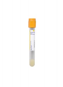
Collection Container:
Gold BD Hemogard™ Serum Separation Tubes (SST)™
Transport Temperature:
Frozen
Volume:
5 mL
Minimum Volume:
2.5 mL
Specimen Type:
Plasma
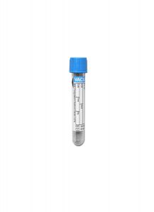
Collection Container:
Light Blue VACUETTE® Sodium Citrate Coagulation Tube
Transport Temperature:
Frozen
Stability
Ambient:
Unacceptable
Refrigerated:
Unacceptable
Frozen:
2 months
Specimen Collection & Handling
Collection of blood by routine venipuncture in a 3.5 mL light blue top tube containing 9:1 ratio of blood to 3.2% trisodium citrate anticoagulant.
Patient Preparation
Discontinue heparin therapy for 2 days prior to collection.
If tests are abnormal, the following tests may be ordered and billed:
Reference Range
Refer to Table 1
Additional Information
Background Information
Antiphospholipid syndrome (APS) is the most common cause of acquired thrombophilia, and the presence of antiphospholipid antibody (APA) is associated with significant morbidity and mortality across diverse patient populations. Both primary and secondary forms of APAs exist, the difference being whether they arise spontaneously or in association with another condition. These antibodies — also known as lupus anticoagulants due to their prevalence in patients with systemic lupus erythematosus — are extremely heterogeneous and are directed against a wide variety of anionic phospholipids, including cardiolipin, ß2 glycoprotein 1 (B2GP1), cell-membrane phosphatidylserine, and many others. Paradoxically, APAs prolong clot-based assays in vitro while predisposing to thrombosis in vivo. In fact, approximately 30% of APA patients will experience thrombosis. A panel of assays is necessary to detect APAs, as no single test presently available is sufficient.
Diagnosis of antiphospholipid syndrome is made by clinicopathologic evaluation. In addition to clinical criteria, such as vascular thrombosis or pregnancy morbidity, repeated laboratory testing of APA is required for the diagnosis because of transient low-level increase of APA in many clinical conditions including infection. The laboratory criteria include positive testing for one of the following on 2 or more occasions, at least 12 weeks apart: 1. lupus anticoagulant; 2. anticardiolipin antibodies (IgG or IgM) in medium or high titer; 3. B2GP1 antibodies (IgG or IgM).
Lupus Anticoagulant (LA) Testing:
Based upon consensus criteria from the International Society for Thrombosis and Haemostasis (ISTH), confirmation of a LA requires that the following criteria are met:
- Performing two or more phospholipid-dependent clotting tests and demonstrating prolongation of at least one test (i.e. aPTT or dilute Russell Viper Venom Test (dRVVT))
- Evidence for inhibitory activity shown by the effect of patient plasma on normal pooled plasma. (i.e. positive mixing study)
- Demonstration of phospholipid-dependence of the inhibitor on a confirmatory test shown by shortening of the clotting time with the addition of more phospholipid
- Exclusion of a co-existing specific factor inhibitor, particularly factor VIII or an anticoagulant drug such as heparin or direct thrombin inhibitor (DTI)
Anticardiolipin Antibody (ACA) IgG, IgM or IgA, and B2GP1 Antibody IgG or IgM Testing:
ACAs recognize a complex of cardiolipin, a naturally found phospholipid, bound to a protein called B2GP1. Complexes of anionic phospholipids and endogenous plasma proteins provide more than one epitope recognized by natural autoantibodies.
An enzyme-linked immunosorbent assay (ELISA) is performed for APA testing. Because the antigen target of ACAs is B2GP1 bound to cardiolipin, B2GP1 antibodies are considered to be more specific than ACA assays.
Clinical Indications
Suspicion for APS in patients with an elevated aPTT, unexplained thrombocytopenia, or a history of arterial and venous thrombosis and/or obstetric complications.
Interpretation
Lupus Anticoagulant (LA)
Tests for LA are interpreted as positive, indeterminate or negative. A narrative interpretation is issued for each patient panel:
Positive:
The panel of tests meets all four diagnostic criteria. If one screening test, one mixing test, and one confirmatory test are positive and there is no evidence for a factor inhibitor or anticoagulant drug effect, the diagnostic criteria for LA are fulfilled.
Indeterminate:
Fewer than four diagnostic criteria are met. If clinical suspicion exists, the patient should be retested at a later date.
Negative:
None of the four diagnostic criteria is met.
Anticardiolipin Antibodies and B2GP1 Antibodies
Tests for ACA and B2GP1 are interpreted as positive, equivocal, or negative. The reference range of each test in the diagnostic panel is shown in Table 1.
Methodology
Laboratory testing for LA consists of a panel of assays (at least two assays on different principles in each criterion) specifically performed together to maximize diagnostic potential.
Test Category
Tests Performed
Screening Tests:
aPTT, aPTT Screen, dRVVT Screen, Hexagonal PL Screen
Four screening tests are performed: the standard laboratory automated aPTT, a more APA-sensitive manual aPTT screen reagent (which contains a different phospholipid composition), the dilute Russell’s viper venom test (dRVVT), a clot-based assay that uses snake venom to activate Factor X directly, and the hexagonal PL screen, which uses a very dilute aPTT reagent to increase sensitivity to phospholipids.
Mixing Studies:
Mixing Study aPTT (immediate and delayed), dRVVT Mix
Patient plasma and normal control plasma are mixed 1:1, and an aPTT and dRVVT test is performed on the mixed sample.
In the presence of an inhibitor in the patient’s plasma, the normal plasma also is affected, and the clotting time will not correct into the normal range. However, if the initial prolonged clotting time was due to a factor deficiency in the patient’s plasma, the normal plasma corrects this deficiency and the resultant clotting time will be normal.
The aPTT mixing study includes a one-hour incubation step to check for more slow-acting specific factor inhibitors
PL Confirmatory Tests:
dRVVT Confirm Ratio, Hexagonal PL Confirm, Platelet Neutralization
Several tests are used to confirm the phospholipid-dependence of an inhibitor:
• The dRVVT confirm ratio is performed by adding PL to plasma and repeating the dRVVT assay. The ratio is calculated by the dRVVT screen/dRVVT confirm.
• The hexagonal phase phospholipid test (STAclot) confirm is performed by adding hexagonal PL to plasma and repeating the hexagonal PL screen. The Delta is calculated by the hexagonal PL screen — the hexagonal PL confirm.
• The platelet neutralization procedure (PNP) uses phospholipid-containing platelet membranes to neutralize the aPTT-prolonging effects of an LA. A PNP test is positive when the prolonged aPTT is shortened by the addition of platelet lysate.
Exclusion Assays:
The presence of other inhibitors must be excluded to confirm the presence of an APA. These include drugs (heparin, DTIs) and specific factor inhibitors (factor VIII is the most common). Tests for each of these are included in the panel, as required per the LA algorithm.
Specific antibodies against cardiolipin and B2GP1 are measured by solid-phase ELISA assay.
Abbreviations:
APTT: Activated partial thromboplastin time
dRVVT: Dilute Russell’s viper venom test
FVIII: Factor VIII
LA: Lupus anticoagulant
PL: Phospholipid
PNP: Platelet neutralization procedure
TT: Thrombin time
Limitations
LAs are heterogeneous in terms of antigenic recognition, and aPTT reagents are variable in terms of phospholipid composition. Thus, variability in detection of LAs may exist between individual reagents, between different panel tests, and/or between laboratories.
Consequently, a normal aPTT cannot definitively exclude the presence of an LA; therefore, if clinical suspicion is high, the full panel may be performed.
Both ACA and B2GP1 APA assays are recommended as using one B2GP1 antibody assay can miss some cases of APA.
Table 1: Reference Ranges of Each Test in the Lupus Anticoagulant Diagnostic Interpretive Panel
Test
Reference Range
aPTT
23.0 – 32.4 sec
PT/INR
8.4 – 13.0 sec / 0.8 – 1.2 sec
TT
<18.6 sec
Mixing Study, Incubated aPTT
Negative
Hexagonal Phase PL Test
Screen:
48.9 – 70.2 sec
Delta:
< 9.0
DRVVT
Screen:
32.7 – 46.7 sec
1:1 Mix:
32.7 – 46.7 sec
Confirm Ratio:
< 1.21
PNP
Negative
ACA IgA
Negative:
< 12 APL
Equivocal:
12 – 40 APL
High Positive:
> 40 APL
ACA IgG
Negative:
< 10 GPL
Equivocal:
10 – 40 GPL
High Positive:
> 40 GPL
ACA IgM
Negative:
< 12 MPL
Equivocal:
12 – 40 MPL
High Positive:
> 40 MPL
B2GP1 Autoabs
IgG:
< 20 Units
IgM:
< 20 Units
Heparin Assay/Factor Xa Inhibition
< 0.10 IU/mL
References
1. Pengo V, Tripodi A, Reber G, et al. Update of the guidelines for lupus anticoagulant detection. J. Thromb Haemost. 2009: 7:1737.
2. Kottke-Marchant K. An Algorithmic Approach to Hemostasis Testing. CAP Press (2008).
3. Miyakis S, Lockshin MD, Atsumi T et al. International consensus statement on an update of the classification criteria for definite antiphospholipid antibody syndrome (APS). J. Thromb Haemost. 2006; 4:295.
4. Moffat KA, Ledford-Kraemer MR, Plumhoff EA et al. Are laboratories following published recommendations for lupus anticoagulant testing? An international evaluation of practices. Thromb Haemost. 2009:101:178.
5. Hoppensteadt D, Walenga J. The relationship between the antiphospholipid syndrome and heparin-induced thrombocytopenia. Hematol Oncol Clin N Am. 2008:22:1.