Test Name
PNH Panel by FCM (PNHPNL)
CPT Codes
88184
88185 (x2)
88187
Methodology
Flow Cytometry (FC)
Turnaround Time
1 – 3 days
Specimen Requirements
Type:
Whole blood
Volume:
4 mL
Minimum Volume:
2 mL
Tube/Container:
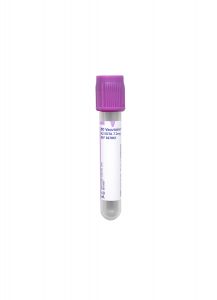
Lavender BD Hemogard™ K2EDTA Tube
Transport Temperature:
Ambient
Stability:
Plasma
Ambient:
48 hours
Refrigerated:
Unacceptable
Frozen:
Unacceptable
Reference Ranges
Negative:
No PNH clone detected
Additional Information
Background Information
Paroxysmal nocturnal hemoglobinuria (PNH) is a clonal stem cell disorder characterized by mutations in the PIGA gene leading to loss of cell surface proteins linked to glycosylphosphatidylinositol (GPI) anchors. Patients affected by PNH display complement-mediated hemolysis, thrombosis, and bone marrow failure, though the clinical presentation is variable. The presence of a PNH clone occurs in classical hemolytic PNH, generally at levels above 1%; however, PNH clones may also be seen in other disorders such as aplastic anemia and myelodysplastic syndrome. Flow cytometric immunophenotypic analysis is the method of choice to detect populations of GPI anchor-deficient cells used in the diagnosis of PNH and to monitor patients with an established diagnosis.1 PNH erythrocyte clones may be divided into those with partial loss of CD59 (PNH Type II Cells) or complete loss of CD59 (PNH Type III Cells).
Cleveland Clinic Laboratories offers high sensitivity flow cytometry testing for paroxysmal nocturnal hemoglobinuria using whole blood. This procedure labels red blood cells (RBC) and white blood cells (WBC) for the detection of GPI-linked surface antigens using monoclonal antibodies and a fluorochrome-labeled bacterial aerolysin (FLAER).2 Through high sensitivity flow cytometry testing, as few as 0.01% PNH cells can be detected.1
Clinical Indications
This test may be useful in the evaluation of patients with intravasuclar hemolysis, unexplained hemolysis, thrombosis with unusual features, or bone marrow failure.
Interpretation
Results are reported as:
- Negative
- Low-level PNH clone positive (.01 – <1%) with percentage
- PNH clone positive (≥1%) with percentage
Limitations
Results of PNH flow cytometry studies must be interpreted in the context of the clinical, laboratory, and histologic findings.
Blood not collected in a K2EDTA tube (or one which has been improperly stored and handled prior to receipt) cannot be processed.
Blood stability limit for PNH testing is 24 hours after the stated draw time. Clinical significance of results on specimens 24-48 hours old should be evaluated in the context of other clinical and laboratory findings.
Blood older than 48 hours from draw time cannot be analyzed.
Methodology
High sensitivity flow cytometry for PNH is performed in accordance with Clinical Cytometry Society recommendations for high sensitivity flow cytometry testing.1
Cells are stained directly with FITC, PE, PE/Cy7, PerCP/Cy5.5, APC, and APC/Cy7-labeled monoclonal antibodies to detect antigens of interest. Antibodies used include CD15, CD24, CD33, CD45, CD59, and glycophorin A. Additionally, FLAER is employed.
For erythrocytes, antibodies to glycophorin A are used to specifically gate red cells, and PNH clones are identified by lack of CD59 expression. PNH erythrocyte clones are divided into those with partial loss of CD59 (PNH Type II Cells) or complete loss of CD59 (PNH Type III Cells).
For granulocytes, CD15 and CD33 are used to specifically gate granulocytes. PNH-type granulocytes are then identified by lack of expression of CD24 and lack of reactivity to FLAER.
References
1. Borowitz MJ, Craig FE, DiGiuseppe JA et al. Guidelines for the Diagnosis and Monitoring of Paroxysmal Nocturnal Hemoglobinuria and Related Disorders by Flow Cytometry. Cytometry B Clin Cytom. 2010; 78B:211-230.
2. Sutherland DR, Kuek N, Davidson J et al. Diagnosing PNH with FLAER and Multiparameter Flow Cytometry. Cytometry B Clin Cytom. 2007; 72B: 167-177.

