October 2018: Changes To COMPC1, COMP3A, & COMP4A Testing

Special Communication

Changes To COMPC1, COMP3A, & COMP4A Testing

Changes effective as of November 5, 2018.

Complement C 1 (COMPC1)

Separate specimens must be submitted when multiple tests are ordered.

Volume:
1 mL, serum

Minimum Volume:
0.25 mL

Collection Container:
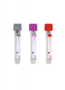
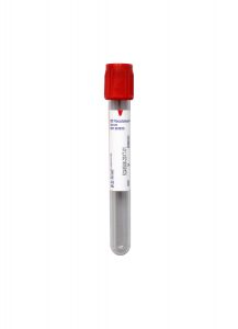
Red BD Hemogard™ Serum Tube (No Additive)

Collection Instructions:
Allow blood to clot for 20 – 60 minutes at room temperature (or 37 °C), then centrifuge.  Aliquot serum into a standard aliquot tube, then freeze the specimen immediately on dry ice or at -70 °C.

Do not draw gel separator tubes.

Transport Temperature:
Critical Frozen – send the frozen specimen via Priority Overnight in a well-insulated container on dry ice.

Thawed specimens or specimens stored at or above -20°C will not be accepted.

Reference Range
• 116,373 – 264,072 units/mL

Complement Component Level 3A (COMP3A)

Separate specimens must be submitted when multiple tests are ordered.

Volume:
1 mL, plasma

Minimum volume:
0.5 mL

Collection Container:
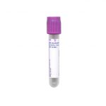
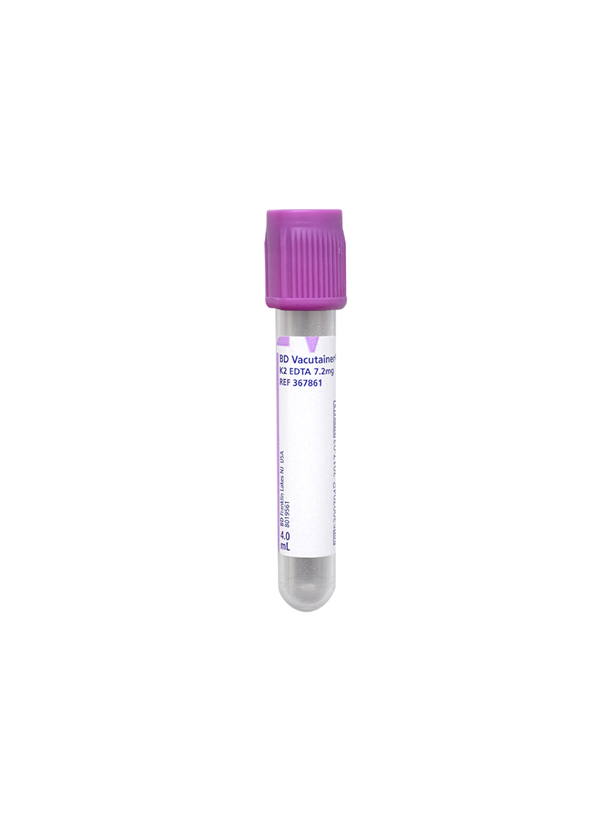
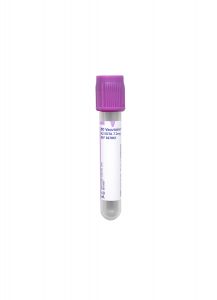
Lavender BD Hemogard™ K2EDTA Tube

Collection Instructions:
Mix well. Centrifuge the specimen at room temperature within 30 minutes of collection, then transfer the plasma to a standard aliquot tube.  Immediately freeze the specimen on dry ice or at -70°C.

Transport Temperature:
Critical Frozen – send the frozen specimen via Priority Overnight in a well-insulated container on dry ice.

Thawed specimens or specimens stored at or above -20°C will not be accepted.

Reference Range
• 0 – 780 ng/mL

Complement Component Level 4A (COMP4A)

Separate specimens must be submitted when multiple tests are ordered.

Volume:
1 mL, plasma

Minimum volume:
0.5 mL

Collection Container:
Lavender BD Hemogard™ K2EDTA Tube

Collection Instructions:
Mix well. Centrifuge at room temperature within 30 minutes of collection, then transfer plasma to a standard aliquot tube and freeze immediately.  Plasma may be frozen at -20°C then transferred to dry ice for shipment within 6 hours or immediately frozen on dry ice/at -70°C or below.

Transport Temperature:
Critical Frozen – send the frozen specimen via Priority Overnight in a well-insulated container on dry ice.

Thawed specimens or specimens stored at room temperature prior to shipment are unacceptable.

Reference Range
• 0 – 2,830 ng/mL

Any additional information will be included in a future Technical Update.  If you have any questions about these changes, please contact Client Services for assistance.

October 2018: Changes to ASPIGG, GAL3, HV6ABS, LIDO, & PROPA Testing

Special Communication

Changes to ASPIGG, GAL3, HV6ABS, LIDO, & PROPA Testing

Effective November 12, 2018.

Aspergillus fumigatus Antibody IgG (ASPIGG)

Previously Aspergillus fumigatus Antibody, IgG by ELISA

Volume:
0.5 mL, serum

Minimum Volume:
0.2 mL

Specimen Container:
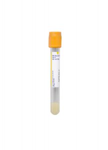
Gold BD Hemogard™ Serum Separation Tubes (SST)™

Galectin-3 (GAL3)

Volume:
1 mL, serum

Specimen Container:
Red BD Hemogard™ Serum Tube (No Additive)

Do not draw Gold Serum Separation Tube (SST) tubes.

Propafenone (PROPA)

Volume:
2 mL, serum

Minimum Volume:
0.7 mL

Specimen Container:
Red BD Hemogard™ Serum Tube (No Additive)

Volume (Alternative):
2 mL, plasma

Minimum Volume:
0.7 mL

Alternative Specimen Container:
Lavender BD Hemogard™ K2EDTA Tube

Effective November 26, 2018.

Herpesvirus 6 Human IgG & IgM Abs (HV6ABS)

Volume:
0.5 mL, serum

Specimen Container:
Red BD Hemogard™ Serum Tube (No Additive)

Transport Temperature:
Ambient (room temperature)

Effective November 27, 2018.

Lidocaine (LIDO)

Volume:
1 mL, plasma

Specimen Container:
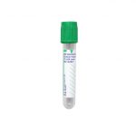
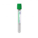
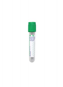
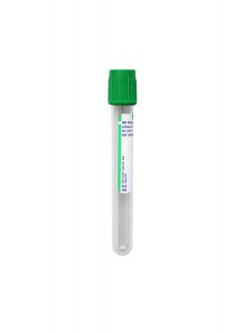
Green Sodium Heparin or Lithium Heparin Tube

Volume (Alternative):
1 mL, serum

Alternative Specimen Container:
Red BD Hemogard™ Serum Tube (No Additive)

Effective November 12, 2018.

Reference Range Changes

• Arsenic, Blood (ASB)
Aspergillus fumigatus Antibody IgG (ASPIGG)
• Eosinophil Cationic Protein (EOSPRO)
• Heavy Metals Screen, Whole Blood (HEVMET)
• Heavy Metals, Urine (UTXM3)
• Heavy Metals with Cadmium, Urine (UTXM4)
• Heavy Metals with Cadmium, Whole Blood (HEVMT4)

• Lead, Urine 24 Hour (ULEADQ)
• Mercury, Blood (MERC2)
• Mercury, Urine 24 Hour (UMERC3)
• Mexiletine (MEX)
• Propafenone (PROPA)*
• Sotalol (SOTAL)*
• Thallium, Urine (UTHAL)

Additional reference range changes:

October 22, 2018
Galactose-alpha-1,3-galactose IgE (13GAL)

November 26, 2018
Herpesvirus 6 Human IgG & IgM Abs* (HV6ABS)

November 27, 2018
Lidocaine (LIDO)

Interfaced clients may need to adjust the test build for Herpesvirus 6 Human IgG & IgM Abs (HV6ABS)Propafenone (PROPA), and Sotalol (SOTAL).

Any additional information will be included in a future Technical Update.  If you have any questions about these changes, please contact Client Services for assistance.
QuantiFERON-TB Gold Plus (QTB-Plus) 4-Tube Assay

Special Communication

QuantiFERON-TB Gold Plus

Effective September 12, 2018.

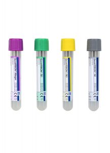
Cleveland Clinic Laboratories will only accept the QuantiFERON-TB Gold Plus (QTB-Plus) 4-Tube Assay for Mycobacterium tuberculosis testing (Normal: INFTBG, INFINC; High Altitude: INFTBP, INTPGP)

New QuantiFERON-TB Gold Plus Collection Tube Kits have a total of four (4) blood collection tubes instead of three.

Regular and High Altitude tubes are available to order from the Supply Storefront.

Please note: Prior to collection, tubes must be stored at 4° to 25°C.

Important Details

The 3-Tube QuantiFERON System will no longer be accepted.

Line draws will no longer be accepted.
•  Venipuncture is the only acceptable draw type.

New order codes will be listed in the Test Directory on September 12, 2018.
• Collection sites, processing, handling, and incubation specifications will remain the same.

All unused 3-Tube QuantiFERON Kits should be returned to our laboratories.
• Tubes can be given to couriers or transport can be arranged through Client Services.

Any additional information will be included in a future Technical Update.  If you have any questions about these changes, please contact Client Services for assistance.
August 2018: Changes to ADRENL & RICKGM Testing

Special Communication

Changes to ADRENL & RICKGM Testing

Adrenal Antibody (ADRENL)

Effective September 10, 2018.

Specimen Requirements

Volume:
2 mL, serum

Minimum Volume:
0.5 mL, serum

Specimen Container:
Red BD Hemogard™ Serum Tube (No Additive)

Do not use Gold Serum Separation Tubes (SST).

Transport:
Refrigerated – send specimen to laboratories with cold packs

Interfaced Clients Only: The ADRENL test build may need to be modified.

Stability

Ambient:
48 hours

Refrigerated:
14 days

Frozen:
30 days

Methodology

Immunofluorescence

Reference Range

Normal Individuals
Negative

Days Performed/Reported

Performed
Wednesday, Friday

Turnaround Time
3–8 days

Special Information

If the Adrenal Antibody screen is positive, then an Adrenal Antibody Titer will be performed at an additional cost.

Clinical Information

Adrenal antibody is detected in patients with autoimmune adrenal disease (e.g., Addison’s disease).

Discontinued: Rickettsia Antibodies, IgG & IgM

Effective September 10, 2018, Rickettsia Antibodies IgG & IgM (RICKGM) will be discontinued.

We suggest ordering Rickettsia rickettsii IgG & IgM Abs (ROCKY) and Rickettsia typhi IgG & IgM Abs (TYPHUS).

Any additional information will be included in a future Technical Update.  If you have any questions about these changes, please contact Client Services for assistance.
Pathology Insights – Lung Granulomas Caused by Fungal Infection with Sanjay Mukhopadhyay, MD

Pathology Insights Video Series

Lung Granulomas Caused by Fungal Infection

Presented by Sanjay Mukhopadhyay, MD

Not all lung nodules are malignant.

In this video, Dr. Mukhopadhyay compares and contrasts two cases of lung granulomas caused by fungal infection. These types of fungal infection cause granulomas in the lung, which can be mistaken for each other.

As part of our educational mission for our clients and communities, Cleveland Clinic Laboratories presents the Pathology Insights video series.
These short videos break down information about interesting pathology cases to better inform doctors, laboratory staff, patients, or anyone interested in the field of pathology. Each episode features important cases, methods, and practices that are personally presented by our staff pathologists.

MGMT Pyrosequencing Methylation Assay for Glioblastoma

Technical Brief

MGMT Pyrosequencing Methylation Assay for Glioblastoma


Test Name

MGMT Methylation (MGMT)

CPT Codes

81287

G0452

Methodology

Polymerase Chain Reaction (PCR)

Pyrosequencing

Turnaround Time

7 days

Specimen Requirements

Type:
Formalin-fixed, paraffin-embedded block

Volume:
1 block

Transport Temperature:
Ambient

Minimal biopsy size 0.5 x 0.5 x 0.5 cm with mainly tumor tissue. Necrosis area should be less than 15%.

Cut 12 charged, unbaked slides, 4um thick. Pre- and post- H&E slides must be examined. Tumor area will be microdissected from USS.

Alternative Specimen

Type:
Formalin-fixed, paraffin-embedded block

Volume:
1 block

Transport Temperature:
Ambient

Minimal biopsy size 0.5 x 0.5 x 0.5 cm with mainly tumor tissue. Necrosis area should be less than 15%.

Cut 8 sections, 10 um thick and place in microcentrifuge tube. Pre- and post- H&E slides must be examined. Tumor area will be microdissected from USS.

Stability

Ambient:
Acceptable

Refrigerated:
Unacceptable

Frozen:
Unacceptable

Reference Range

Positive for MGMT methylation: ≥ of 10% of any CpG island or average of all 5 CpG islands.

Negative for MGMT methylation: >10% of methylation or average of all CpG islands analyzed.

Background Information

Glioblastoma is the most common and most aggressive malignant primary brain tumor. While occurring in only two to three cases per 100,000 people in North America, glioblastoma represents 52% of all functional tissue brain tumor cases and 20% of all intracranial tumors. Prognosis for those diagnosed with glioblastoma is poor, with a median survival time of about 14 months.1

Patients with glioblastoma can be treated with alkylating agents, such as Temador® (temozolomide). Epigenetic silencing of the MGMT (O6-methylguanine-DNA methyltransferase) DNA-repair gene by promoter methylation compromises DNA repair and has been associated with longer survival in patients with glioblastoma who receive temozolomide.2,3

Temozolomide kills tumor cells by producing cross-links between DNA strands and inhibiting DNA replication. The most common alkylation site is the O6 position of guanine. O6-methylguanine DNA methyltransferase (MGMT) is a DNA repair protein that reverses such DNA alkylation and confers chemoresistance by repairing DNA damage. Temozolomide seems to work by sensitizing the tumor cells to radiation.4

Recent clinical studies confirm that the presence of MGMT promoter methylation in tumor samples corresponds to an increased likelihood that tumor cells would be responsive to temozolomide.3,4,5,6 If the promotor was methylated, temozolomide was more effective. It is estimated that approximately 40 to 50% of glioblastoma tumors exhibit MGMT gene methylation, which correlates significantly with reduced DNA damage repair induced by alkylating agents and significantly enhanced chemosensitivity.4

According to recent clinical trials, glioblastoma patients with MGMT methylation respond to temozolomide two to three times better than those lacking of MGMT methylation. Prolonged overall and progression-free survival at 24 months was 80% for those with MGMT methylation vs. 20% for those lacking MGMT methylation.

Diagnostic MGMT testing requires sufficient and optimally preserved tumor tissue. Cleveland Clinic’s MGMT methylation assay is a new, quantitative MSP test to detect the methylation status of brain tissue that has undergone thorough clinical evaluation. Our protocol calls for a minimal tissue sample size of ½-centimeter, which is much smaller than other laboratories that typically require at least a minimum 1-centimeter sample size. The best results are obtained with cryopreserved tumor specimens.

Clinical Indications

For patients diagnosed with glioblastoma to determine if a methylated MGMT promoter is present, which is a favorable prognostic indicator for temozolomide treatment. Individuals without a methylated MGMT promoter do not have such a benefit.

MGMT “silence” is the most significant guide for the treatment of glioblastoma. This assay is to validate the methylation status of the MGMT gene.

Methodology

Pyrosequencing technology, which is based on the principle of sequencing by synthesis, provides quantitative data in sequence context within minutes. Real-time sequence information is highly suitable for quantification of CpG methylation. We have validated pyrosequencing-based assay in detection of MGMT methylation in paraffin-embedded biopsy tissue specimens. With 10% average methylation as a cutoff, MGMT promoter methylation was detected in glioblastoma, but not detected in non-neoplastic brain tissue. The analytical sensitivity of the assay is 5% of target cells harboring MGMT methylation.

Diagnostic MGMT testing requires sufficient and optimally preserved tumor tissue. The biopsy should be at least 0.5 cm in size and necrosis should be less than 15%. Both frozen and paraffin-embedded tissue are suitable for the pyrosequencing-based MGMT methylation assay.

Interpretation

Positive for MGMT methylation:
Equal or greater than 10% of methylation in any CpG island or in average of all CpG islands analyzed.

Negative for MGMT methylation:
Less than 10% of any CpG island or in average of all CpG islands analyzed.

References

1. Van Meir EG, et al. “Exciting New Advances in NeuroOncology: The Avenue to a Cure for Malignant Glioma”. CA: A Cancer Journal for Clinicians. 60 (3):166–93. doi:10.3322/caac.20069. PMC 2888474. PMID 20445000. 2010.

2. Stupp R, et al. Radiotherapy plus Concomitant and Adjuvant Temozolomide for Glioblastoma. N Engl J Med. 352:987-996.

3. Hegi M, et al. MGMT Gene Silencing and Benefit from Temozolomide in Glioblastoma. N Engl J Med. 2005;352;10:997-1003.

4. Chamberlain Marc C, et al. “Early necrosis following concurrent Temodar and radiotherapy in patients with glioblastoma”. Journal of Neuro-Oncology. 82(1):81-3. doi:10.1007/s11060-006-9241-y. PMID 16944309.

5. Vlassenbroeck I, et al. Validation of Real-Time MethylationSpecific PCR to Determine O(6)-Methylguanine-DNA Methyl-transferase Gene Promoter Methylation in Glioma. J. Molecular Diagnostics. 2008;10;4:332-337.

6. Brandes A, et al. MGMT Promoter Methylation Status Can Predict the Incidence and Outcome of Pseudoprogression After Concomitant Radiochemotheraphy in Newly Diagnosed Glioblastoma Patients. J. Clinical Oncology. 2008; 26;13:2192-2197.

June 2018: Changes to Cyanide, Blood (CYANID) Testing

Special Communication

Changes to Cyanide, Blood (CYANID) Testing

Effective August 15, 2018.

Specimen Requirements

Volume:
4 mL, whole blood

Minimum Volume:
3 mL, whole blood

Specimen Container (Primary):
Green Sodium Heparin Tube

Specimen Container (Primary):
Green Lithium Heparin Tube

Alternative Container:
Lavender BD Hemogard™ K2EDTA Tube

Serum or plasma specimens are unacceptable.

Transport:
Ambient (room) temperature

Patient Preparation:
Timing of specimen collection is dependent on the time of exposure; test upon presentation to hospital.

Stability

Ambient
72 hours, if tightly-capped

Refrigerated
Unacceptable

Frozen
Unacceptable

Methodology

Quantitative Colorimetric

Reference Range

Non-smoker: < 20 μg/dL

Smoker: < 40 μg/dL

Days Performed/Reported

Performed
Sunday, Wednesday, and Friday

Turnaround Time
2–6 days

Special Information

Timing of specimen collection is dependent on the time of exposure; test upon presentation to hospital.

This test is New York DOH approved.

Frozen or refrigerated specimens will be rejected.

Clotted or hemolyzed specimens will be rejected.

Serum or plasma specimens are unacceptable.

Clinical Information

This test is used to monitor cyanide exposure. Cyanide poisoning can cause hypoxia, dizziness, weakness, and mental and motor impairment.

Elevated cyanide concentrations rarely indicate toxicity for patients on nitroprusside therapy. Thiocyanate should be monitored in patients on nitroprusside therapy for potential toxicity. Toxicity may occur with long-term nitroprusside use (longer than 7–14 days with normal renal function, and 3-6 days with renal impairment at greater than 2 μg/kg/min infusion rates).

Thiocyanate levels may be monitored on an every-other-day basis to assess potential thiocyanate toxicity and to indicate possible adjustments in dosage.

Any additional information will be included in a future Technical Update.  If you have any questions about these changes, please contact Client Services for assistance.
Pathology Insights – Lung Cancer or Metastasis to Lung? with Sanjay Mukhopadhyay, MD

Pathology Insights Video Series

Lung Cancer or Metastasis to Lung?

Presented by Sanjay Mukhopadhyay, MD

Can metastases to the lung occur from cancers that were treated several years ago?  Dr. Mukhopadhyay discusses.

As part of our educational mission for our clients and communities, Cleveland Clinic Laboratories presents the Pathology Insights video series.
These short videos break down information about interesting pathology cases to better inform doctors, laboratory staff, patients, or anyone interested in the field of pathology. Each episode features important cases, methods, and practices that are personally presented by our staff pathologists.

BCR / ABL p210 Quantitative RT-PCR Assay

Technical Brief

BCR / ABL p210 Quantitative RT-PCR Assay with International Scale Reporting for Minimal Residual Disease in Chronic Myeloid Leukemia


Test Name

BCR / ABL p210 RT-PCR, quantitative (BCRPCR)

CPT Code

81206
G0452

Methodology

Reverse Transcription/Polymerase Chain Reaction (RT/PCR)

Turnaround Time

5 – 7 days

Specimen Requirements

Clients should ship specimens as “Priority Overnight” to ensure specimen stability.

Do not ship on Fridays or the day preceding a holiday.

Specimen Type:
Whole blood

Volume:
10 mL

Minimum Volume:
4 mL

Collection Container:
Lavender BD Hemogard™ K2EDTA Tube

Transport Temperature:
Ambient

Clearly indicate specimen type on the label.

Alternative Specimen

Specimen Type:
Bone marrow

Volume:
5 mL

Minimum Volume:
2 mL

Collection Container:
Lavender BD Hemogard™ K2EDTA Tube

Transport Temperature:
Ambient

Clearly indicate specimen type on the label.

Stability

Ambient:
48 hours

Refrigerated:
7 days

Ambient:
Unacceptable

Reference Range

BCR ABL p210 transcripts not detected

Additional Information

Background Information

A translocation between chromosomes 9 and 22, resulting in the formation of a BCR / ABL fusion transcript, has long been recognized as a hallmark of chronic myeloid leukemia (CML). Although the breakpoints in BCR and ABL are variable, in >95% of cases of CML, the translocation results in production of the p210 isoform of the fusion protein (e13a2 or e14a2 fusion genes).

Modern therapy for CML, including tyrosine kinase inhibitors, has resulted in effective therapeutic options for CML patients. With this advance in treatment has come the need for effective monitoring for the presence of minimal residual disease (MRD) and the ability to recognize disease progression at an early stage. Techniques such as fluorescence in situ
hybridization (FISH) and metaphase cytogenetics provide valuable information at the time of initial diagnosis of CML and metaphase cytogenetics is helpful for identifying the emergence of additional chromosomal abnormalities, however, neither of these techniques are sufficiently sensitive to monitor MRD.[1],[2] For this reason, a BCR ABL p210 quantitative real-time reverse transcriptase polymerase chain reaction (qRT-PCR) assay was developed, validated, and implemented in the Department of Molecular Pathology.

To facilitate comparison of quantitative RT-PCR results between laboratories and platforms, International Scale reference materials were established by the World Health Organization.[3] Results of this assay are now reported on the International Scale.

Clinical Indications

Quantitative detection of BCR/ABL p210 transcripts (e13a2 or e14a2) in patients with CML.

Methodology

Assay sensitivity was established using RNA extracted from K562 cultured cells suspended in normal buffy coat. This assay successfully detects p210 BCR ABL transcripts in RNA extracted from one K562 cell in 100,000 peripheral blood leukocytes from a normal individual.

Interpretation

Results are reported as the percentage ratio of fusion gene transcripts to wild-type ABL transcripts (% BCRABL ABL).

Results are also converted to the International Scale. A BCRABL ABL value of 0.1% on the International Scale represents a major molecular response by consensus criteria.

References

1. Hughes T, Deininger M, Hochhaus A et al. Monitoring CML patients responding to treatment with tyrosine kinase inhibitors: review and recommendations for harmonizing current methodology for detecting BCRABL transcripts and kinase domain mutations and for expressing results. Blood. 2006;108:28-37.

2. Druker BJ, Guilhot F, O’Brien SG et al. Five-year follow-up of patients receiving imatinib for chronic myelogenous leukemia. N Engl J Med. 2006;355:2408-2417.

3. White HE, Matejschuk P, Rigsby P, et al. Establishment of the first World Health Organization International Genetic Reference Panel for quantification of BCRABL mRNA. Blood 2010;116:e111-7.

April 2018: Changes to Chemistry Tests

Special Communication

Changes to Chemistry Tests

Changes effective as of April 17, 2018

Tests

6-Monoacetylmorphine (6-MAM) Confirmation, Urine (U6AMCO)

Amphetamine Confirmation, Urine (UAMPC)

Benzodiazepines Conf, Urine (UBENZC)

Benzoylecgonine Confirmation/Quantitation (BECGO)

Buprenorphine Quant, Urine (UQNTBU)

Cannabinoid Confirmation, Urine (UTHCC)

Cocaine Confirmation, Urine (UCOCC)

Fentanyl and Metabolite, Urine (UFENT)

Methadone Quantitation, Urine (UQMET)

Opiate Confirmation, Urine (OPICON)

Oxycodone Confirmation, Urine (UOXYCC)

Quantitative Pain Panel, Urine (UQNTPP)

Tramadol and Metabolite, Quantitation (TRAQNT)

Build / Reference Range

Specimen Validity pH, Urine:
4.5 – 8.0

Specimen Validity Specific Gravity, Urine:
1.002 – 1.030

Specimen Validity Creatinine, Urine:

18–99 Years (Male):
46.8 – 314.5 mg/dL

18–99 Years (Female):
42.2 – 237.9 mg/dL

Specimen Validity Nitrites, Urine:
≤ 50 mg/L

Specimen Validity Oxidants, Urine:
< 200 mg/L

Specimen Validity Chromate, Urine:
< 50 mg/L

Oxycodone

In addition to build and special/clinical information changes, the following tests will also have an adjustment to the reference range for Oxycodone:

Tests

Opiate Confirmation, Urine (OPICON)

Oxycodone Confirmation, Urine (UOXYCC)

Quantitative Pain Panel, Urine (UQNTPP)

Reference Range

Oxycodone, Urine:
< 10 ng/mL

Special Information

Specimen Validity Testing (SVT) analytes have a stability of up to 5 days at 2 – 8 °C, as stated in the Instructions for Use (IFU).

Based on the laboratory’s studies, this stability is based on samples that have reached an equilibrium.

  • Introduction of oxidants to a urine sample will not be stable until an equilibrium is achieved.
  • Time to reach equilibrium will vary dependent on the type and amount of oxidant used as an adulterant.

Clinical Information

For Specimen Validity Interpretations, the following rules are applied:

Diluted

Creatinine: ≥ 2 mg/dL but < 20 mg/dL

Specific Gravity: > 1.0010 but < 1.0030

Substituted

Creatinine: < 2 mg/dL

Specific Gravity: < 1.0010 or ≥ 1.0200

Adulterated

pH: < 3.0 or ≥ 11.0

or Nitrites: ≥ 500 mg/L

or Oxidants: ≥ 200 mg/L

or Chromate: ≥ 50 mg/L

Any additional information will be included in a future Technical Update.  If you have any questions about these changes, please contact Client Services for assistance.