Antibody Testing for COVID-19: Overview & FAQs

CAP Statement on the Current Role of Serologic Testing for SARS-CoV-2

May 13, 2020 | From the College of American Pathologists

Understanding COVID-19 & Antibody Testing

May 7, 2020 | From Cleveland Clinic’s Health Essentials:

We’ve accepted the harsh realities of COVID-19 and we’ve adjusted our lives accordingly. And yet, we still have so many unanswered questions like why has COVID-19 taken such a detrimental toll, and where are we in the process of finding a cure?

To get a better idea of where science is headed, Cleveland Clinic CEO and President Tom Mihaljevic, MD interviews Serpil Erzurum, MD, Chair of the Lerner Research Institute.

COVID-19 Serologic Testing: FAQS and Caveats

April 29, 2020 | From the Cleveland Clinic Journal of Medicine’s COVID-19 Curbside Consults:

There has been an immense amount of tangential discussion regarding the potential usefulness of serologic testing for COVID-19 recently. Although serologic testing has never been routinely used for diagnosing infections with “respiratory viruses”, such as influenzae, parainfluenzae, respiratory syncytial viruses, adenoviruses, or metapneumovirus, questioning its usefulness during the current outbreak has relevance.

That being said, during global epidemics of SARS, MERS, and H1N1 influenza, serology was never used in routine diagnostics. However, given the pandemic status of COVID-19 and the shortage of nucleic acid detection kits and/or swabs in certain areas, it raises the prospect of resorting to serology as an alternative to directly testing for the presence of the virus. The Infectious Diseases Society of America (IDAS) has recently issued a clear statement on COVID-19 serology.

Kamran Kadkhoda, PhD

Kamran Kadkhoda, PhD
Medical Director, Immunopathology

Respiratory Virus Testing

Updated August 2024

Respiratory Virus Testing

Cleveland Clinic Laboratories offers several respiratory testing services to healthcare organizations & providers.

SARS-CoV-2 (COVID-19), influenza A/B, and respiratory syncytial virus (RSV) testing is available for the qualitative detection of these viruses in respiratory specimens from individuals with appropriate signs, symptoms, and risk factors.

All three viruses are expected to be in circulation during the 2024-2025 respiratory virus season.

Respiratory Virus Testing Overview

Test Name

COVID, Influenza A/B, & RSV NAAT, Routine (CVFLRS)

Turnaround Time

24 hours from the time of receipt by the laboratory*
Testing is performed 24 hours a day, 7 days a week

Specimen Requirements – Upper Respiratory

Type:
(1) Nasopharyngeal (NP) Swab – preferred 

Volume:
3 mL

Specimen Container:
Universal Transport Media (UTM)

Accepted Alternatives

Type:
• (1) Nasopharyngeal (NP) swab
• (1) Nasal (anterior nares) swab

Volume:
Swab

Transport Media:
• Universal Transport Media (UTM)
• eSwab
• Saline (sterile)
• Viral Transport Media (VTM), including M4RT, M5, or M6

Specimen Requirements – Lower Respiratory

Type:
• Aspirate, tracheal
• Bronchoalveolar Lavage (BAL)
• Sputum

Volume:
1 mL

Transport Container:
Sterile container

Transport Temperature

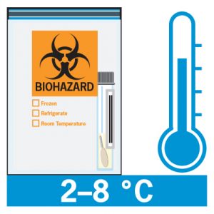
Refrigerated; transport to the laboratories on cold packs or wet ice.

Stability

Ambient:
Unacceptable

Refrigerated (2-8ºC):
96 hours

Frozen:
Up to 30 days

Rejection Criteria

Swabs used for Group A Strep testing are not acceptable.

Expanded Respiratory Pathogen Panel Test Overview

This test is for immunocompromised patients, severely ill adults who will be hospitalized and have evidence of a lower respiratory tract infection the etiology of which has not been determined, and children who are going to be admitted that are being evaluated for a suspected respiratory viral infection.

This panel should rarely be ordered in the outpatient setting, and if ordered, preauthorization should be strongly considered. The test is very expensive and if not covered by insurance the patient will incur a substantial charge.

Test Name

Expanded Respiratory Pathogen Panel by PCR (with COVID), Routine (RPPCR)

Turnaround Time

2 – 3 days

Specimen Requirements – Upper Respiratory

Type:
(1) Nasopharyngeal (NP) Swab – preferred 

Volume:
3 mL

Specimen Container:
Universal Transport Media (UTM)

Accepted Alternatives

Type:
• (1) Nasopharyngeal (NP) swab
• (1) Nasal (anterior nares) swab

Volume:
Swab

Transport Media:
• Universal Transport Media (UTM)
• eSwab
• Saline (sterile)
• Viral Transport Media (VTM), including M4RT, M5, or M6

Specimen Requirements – Lower Respiratory

Type:
• Aspirate, tracheal
• Bronchoalveolar Lavage (BAL)
• Sputum

Volume:
1 mL

Transport Container:
Sterile container

If aliquotting is necessary, sterile aliquot tubes must be used. Do not dilute with UTM or VTM.

Transport Temperature

Refrigerated; transport to the laboratories on cold packs or wet ice.

Stability, Upper Resp.

Ambient:
Unacceptable

Refrigerated (2-8ºC):
3 days

Frozen:
Up to 30 days

Stability, Lower Resp.

Ambient:
Unacceptable

Refrigerated (2-8ºC):
7 days

Frozen:
Up to 30 days

Rejection Criteria

Swabs used for Group A Strep testing are not acceptable.

Ready to Place an Order?

Download and complete a Respiratory Virus Testing Requisition (required).

Need to Set Up an Account?

Request support from a regional Account Manager or contact Client Services for general inquiries.

Specimen Collection Instructions

You will need:

Personal protective equipment (PPE)

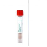
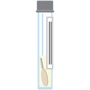
(1) tube of transport medium

– Universal Transport Media (UTM)
– Viral Transport Media (VTM)
– Saline solution

(1) swab

Patient identification labels

Before you begin:

• Confirm the patient’s name and date of birth.

• Wash your hands, then put on personal protective equipment, including gloves.

If you are having difficulties obtaining appropriate COVID-19 specimen collection and transport supplies, please contact your CCL Account Manager.

Nasopharyngeal (NP) Swabs

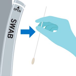
Step 1:
Carefully remove the swab from the packaging without touching the tip.

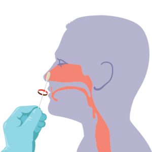
Step 2a for nasopharyngeal (NP) swabs:

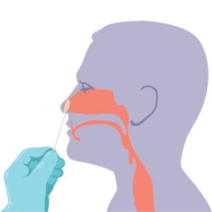
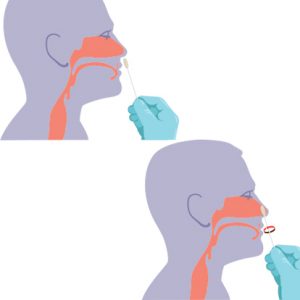
– Gently insert the NP swab along the nasal septum just above the floor of the passage to the nasopharynx until resistance is met.

– Rotate the swab gently against the nasopharyngeal mucosa for 10 – 15 seconds, then remove.

Step 3:
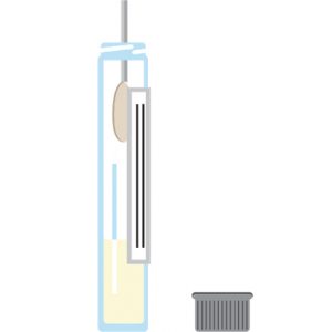
Remove the cap from the tube, then insert the swab all the way to the bottom of the vial.

Step 4:
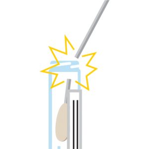
While holding the tube away from your face, bend the swab in the vial at a 180-degree angle to break at the marked breakpoint.

Step 5:
Close the tube tightly. If possible, wrap the cap with Parafilm.

Any leaking specimens will be rejected.

Step 6:
Label specimen with the printed label or write the patient’s first & last name, date of birth, and the date, time, and site of collection (e.g., NP) on the tube.

Step 7:
Double-bag the specimen, place on wet ice, then refrigerate.

Step 8:
Complete a Coronavirus 2019 (COVID) Test Requisition.

Step 9:
Place the completed requisition in the pocket of the specimen bag.

Do not place any paperwork in the part of the bag containing the specimen.

Step 10:
For the fastest turnaround time, deliver the specimen directly to the Main Campus LL Building Specimen Receiving Area:

Cleveland Clinic Laboratories
10300 Carnegie Avenue
Cleveland, OH 44106

Need a COVID-19 Test Requisition? 

Contact your CCL Account Manager or Client Services.

Nasal (Anterior Nares) Swabs

Specimen Collection:

Step 1:Carefully remove the swab from the packaging without touching the tip.

Step 2:

Position the patient’s head slightly back, then insert the swab into their LEFT nostril (less than one inch) and gently push until a slight resistance is met.

Step 3:

Rotate the swab against the inside of the patient’s nostril four times for 10-15 seconds.

Step 4:
Remove the swab, then repeat Steps 2 & 3 in the RIGHT nostril with the same swab.

Remove the swab from the patient’s nose when finished.

Preparing for Transport:

Step 5:

While holding the swab, open the tube and set the cap down with the threads facing up.

Insert the swab into the tube until the breakpoint is level with the tube opening.

Step 6:

While holding the tube away from your face, bend the swab until it snaps at the break point.

You may need to gently rotate the swab shaft to complete the breakage.

Step 7:

After discarding the remaining swab shaft into an approved disposal container, screw the cap back onto the tube.

Apply the test label or write the patient’s information onto the tube.

Step 8:

Place the specimen into a biohazard bag, then insert any lab paperwork into the outside pouch.

Refrigerate the specimen during storage and transport to the lab.

If you have any questions, please contact your CCL Account Manager or Client Services for assistance.

Alpha-1 Antitrypsin (SERPINA1) Targeted Genotyping

Technical Brief:

Alpha-1 Antitrypsin (SERPINA1) Targeted Genotyping


Test Name

Alpha-1 Antitrypsin (SERPINA1) Targeted Genotyping (HA1AT)

CPT Codes

81332

Turnaround Time

7 days

Specimen Requirements

Type:
Peripheral blood

Volume:
4 mL

Minimum Volume:
1 mL

Specimen Container:
Lavender BD Hemogard™ K2EDTA Tube

Transport Temperature:
Ambient

Stability

Ambient:
48 hours

Refrigerated:
7 days

Frozen:
Unacceptable

Methodology

High Resolution Melt Analysis
Real-Time Polymerase Chain Reaction (RT-PCR)
Fluorescence Monitoring

Background Information

Alpha-1 antitrypsin deficiency (AATD) (OMIM#613490) is one of the most commonly inherited metabolic disorders in people of northern European ancestry, occurring in one in 3000-5000 individuals, and also occurs at lower frequencies in people from other regions. AATD predisposes an individual to chronic obstructive pulmonary disease (COPD), liver disease, panniculitis and C-ANCA-positive vasculitis. AATD is caused by pathogenic variants in SERPINA1 (RefSeq NM_001127701.0; GRCh38/hg38), the gene that encodes alpha-1 antitrypsin (AAT). Alpha-1 antitrypsin is an inhibitor of neutrophil elastase. Excess neutrophil elastase can destroy the alveolar walls of the lung, causing emphysema. Pathogenic variants in SERPINA1 can also cause accumulation of abnormal proteins in hepatocytes leading to chronic liver disease. AATD is inherited as an autosomal recessive condition and more than 150 variants in SERPINA1 have been described to date.

Alleles in AATD are named with the prefix PI* for “protease inhibitor,” another name for the AAT protein. Many pathogenic variants in SERPINA1 result in structurally abnormal AAT protein, which impairs secretion and results in plasma deficiency. The majority of patients with AATD have the PI*S (c.863A>T, p.Glu288Val, g.94380925) or PI*Z (c.1096G>A, p.Glu366Lys, g.94378610) alleles. The PI*S allele causes a structurally abnormal AAT with mild functional impact and low disease risk, unless combined with other pathogenic alleles. The PI*Z allele also encodes a structurally abnormal form of AAT with more severe dysfunction associated with higher risk of lung and liver disease. Approximately 95% of individuals with clinical manifestations of AATD have the PI*ZZ genotype. Other rare variants of SERPINA1 exist and can also cause lung and liver disease. The PI*F allele (c.739C>T, p.Arg247Cys, g.94381049) results in a quantitatively normal but functionally abnormal AAT, causing decreased binding to neutrophil elastase. Individuals with this variant may have normal AAT levels but have an increased risk for lung disease, especially when inherited along with PI*Z allele. The PI*I allele (c.187C>T, p.Arg63Cys, g.94383051), similar to the PI*S allele, causes some structural abnormality in AAT with mild functional impact. However, when inherited along with the PI*Z allele, PI*I confers a higher risk of lung and liver disease.

Serum alpha-1 antitrypsin levels typically correlate with the SERPINA1 genotypes as shown in Table 1. However, AAT levels can be significantly elevated in some clinical circumstances, which could mask AATD. Serum levels in patients with acute inflammation, cancer, non-AATD related liver disease, pregnancy, estrogen therapy, or blood transfusions may be discordant from genotype results.

Table 1

Genotype

Expected AAT Levels
(based on Bornhorst 2003 data)

Disease Risks

PI*MM

102-254 mg/dL

Standard lab reference range:
90-200 mg/dL (per Dati 1996)

No increased disease risks

PI*MS

86-218 mg/dL

No increased disease risks

PI*MZ

62-151 mg/dL

No increased disease risks

PI*MF

102-254 mg/dL

No increased disease risks

PI*MI

86-218 mg/dL

No increased disease risks

PI*SS

43-154 mg/dL

Possible pulmonary disease risk

PI*SZ

38-108 mg/dL

Possible pulmonary and hepatic disease risks

PI*ZZ

<52 mg/dL

Pulmonary and hepatic disease risks, risk of other AATD-related conditions (e.g., panniculitis)

PI*FS

86-218 mg/dL

Pulmonary disease risk

PI*IS

43-154 mg/dL

Possible pulmonary disease risk

PI*FZ

38-151 mg/dL

Pulmonary disease risk, possible hepatic disease risk

PI*IZ

38-108 mg/dL

Possible pulmonary and hepatic disease risks

PI*FF

102-254 mg/dL

Pulmonary disease risk

Clinical Indications

According to available guidelines:

Diagnostic testing is recommended for:

  • Adults with emphysema, COPD, or asthma that is incompletely responsive to bronchodilators
  • Individuals with unexplained liver disease
  • Asymptomatic individuals with persistent obstruction on pulmonary function tests with identifiable risk factors
  • Adults with necrotizing panniculitis
  • Siblings of adults with AATD

Testing may be considered for:

  • Adults with bronchiectasis without clear risk factors for bronchiectasis
  • Adolescents with persistent airflow obstruction
  • Asymptomatic individuals with persistent obstruction on pulmonary function tests with no identifiable risk factors
  • Adults with C-ANCA-positive vasculitis
  • Parents or children of adults with AATD
  • Screening for individuals >11 years of age in areas of AATD prevalence or in areas of high smoking rates

Methodology

Targeted variant analysis is performed using LightMix® and LightSNiP® melt curve technology (TIB MOLBIOL) on the LightCycler480 II (Roche) to identify four alleles of the SERPINA1 gene, PI*S, PI*F, PI*I and PI*Z, which combine to create the genotypes listed in Table 1.

Genomic DNA is isolated from the peripheral blood and four regions containing the variants of interest are amplified by PCR. Following PCR, the amplified DNA sequences are subjected to a temperature gradient, allowing fluorescently labeled probes targeted to each variant to bind to the double stranded DNA sequence and fluoresce. As the temperature increases, the probes dissociate, or “melt”, at a specific temperature based on the nucleotide sequence of the probe and fluorescence decreases. Measurement of fluorescence throughout the gradient allows the specific temperature at which melting occurs to be recorded. Mismatches between the probe and the DNA sequence cause the probe/DNA hybrid to be less stable and the probe to melt at a lower temperature, allowing discrimination between variant and normal.

Interpretation

This laboratory-developed test will not detect other mutations that may cause AATD.

Uncommon variants or polymorphisms in the regions of interest may affect the binding of LightMix® or LightSNiP® probes and may result in a false negative, false positive, or indeterminate result.

Absence of the S, Z, F, and I alleles is consistent with (but does not confirm) the normal, aka wild type, PI*MM genotype.

Importantly, there are over 100 known rare variants of SERPINA1 that are not detected in 201306.081 (1.20 rev) by this PCR test. Therefore, the correlation of the genotype with the patient’s serum alpha-1 antitrypsin level and clinical manifestations is strongly recommended.

When discrepancies exist between the enzyme level and targeted genotype results (e.g., the serum AAT level is low but no abnormal allele is identified by PCR), sequencing of the coding regions of the SERPINA1 gene to identify rare mutations should be considered.

References

1. Bornhorst JA, Greene DN, Ashwood ER, Grenache DG. α1-Antitrypsin phenotypes and associated serum protein concentrations in a large clinical population. Chest. 2013;143:1000-8.

2. Dati F, Schumann G, Thomas L, Aguzzi F, et al. Consensus of a group of professional societies and diagnostic companies on guidelines for interim reference ranges for 14 proteins in serum based on the standardization against the IFCC/BCR/CAP reference material (CRM 70), Eur J Clin Chem Biochem. 1996;34:517-520.

3. Rodriguez-Frias F, Miravitlles M, Vidal R, Camos S, Jardi R. Rare alpha-1-antitrypsin variants: are they really so rare? Ther Adv Respir Dis. 2012 Apr;6(2):79-85.

4. Sandhaus RA, Turino G, Brantly ML, Campos M, Cross CE, Goodman K, Hogarth DK, Knight SL, Stocks JM, Stoller JK, Strange C, Teckman J. “The Diagnosis and Management of Alpha-1 Antitrypsin Deficiency in the Adult.” Chronic Obstr Pulm Dis. 2016 Jun 6;3(3):668-682.

5. Silverman EK, Sandhaus RA. Clinical practice. Alpha 1-antitrypsin deficiency. N Engl J Med. 2009;360(26):2749-57.

6. Sinden NJ, Koura F, Stockley RA. The significance of the F variant of alpha-1 antitrypsin and unique case report of a PiFF homozygote. BMC Pulm Med. 2014 Aug 7;14:132.

7. Stoller JK, Aboussouan LS. A review of α1-antitrypsin deficiency. Am J Respir Crit Care Med. 2012;185(3):246-59.

8. Stoller JK, Lacbawan FL, Aboussouan LS. Alpha-1 Antitrypsin Deficiency. 2006 Oct 27 [Updated 2017 January 19]. In Adam MP, Ardinger HH, Pagon RA, et al., editors. GeneReviews. [Internet]. Seattle (WA): University of Washington, Seattle; 1993-2018. Available from: http://www.ncbi.nlm.nih.gov/books/NBK1519/

Thyroid Stimulating Immunoglobulins (TSI) Assay

Technical Brief

Thyroid Stimulating Immunoglobulins (TSI) Assay


Test Name

Thyroid Stimulating Immunoglobulin (TSIGIM)

CPT Code

84445

Methodology

Chemiluminescent Immunoassay (CLIA)

Turnaround Time

1 – 4 days

Specimen Requirements

Type:
Serum

Volume:
0.5 mL

Minimum Volume:
0.35 mL

Specimen Container:
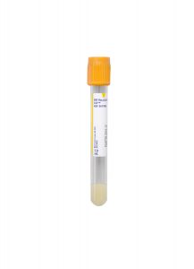
Gold BD Hemogard™ Serum Separation Tubes (SST)™

Transport Temperature:
Refrigerated

Submitting the minimum volume will not allow for repeat testing or add-ons.

Alternative Specimen

Type:
Plasma

Volume:
0.5 mL

Minimum Volume:
0.35 mL

Specimen Container:
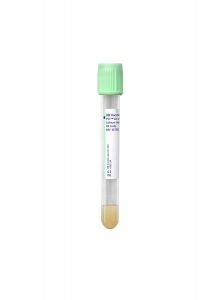
Light Green Lithium Heparin Plasma Separator Tube (PST)

Transport Temperature:
Refrigerated

Submitting the minimum volume will not allow for repeat testing or add-ons.

Stability

Ambient: 
24 hours

Refrigerated:
7 days

Frozen:
1 year (≤ -20°C)

Reference Range

<0.55 IU/L

Linear Range

0.10–40.00 IU/L

Clinical Information

The Thyroid Stimulating Immunoglobulin test is used as an aid in diagnosis of autoimmune hyperthyroidism, especially in patients with Grave’s orbitopathy and dermopathy. Low positive TSH receptor stimulating antibody levels may occasionally be found in patients with autoimmune hypothyroidism. Clinical correlation is required.

Background Information

Grave’s disease (GD) is the most common cause of hyperthyroidism. The clinical manifestations are protean and, to some extent, shared with those of Hashimoto thyroiditis (HT), which is a type of autoimmune hypothyroidism. The shared manifestations may include: muscle weakness, menstrual disturbances, hair and skin changes, decreased concentration, fatigue, goiter, and depression.

The autoimmune nature of Grave’s disease was established when the so-called long-activating thyroid stimulator (LATS) in sera from patients with Grave’s disease was able to induce hyperthyroidism in experimental animals. LATS were later found to be thyroid-stimulating hormone receptor (TSHR)-stimulating antibodies.

TSHR is a membrane receptor that belongs to a superfamily that contains hormone receptors for LH, FSH, and HCG. The extracellular domain (ectodomain, subunit A) of TSHR has several leucine-rich repeats (LRDs) that create a binding site for thyroid-stimulating hormones (TSH). Anti-TSHR stimulating and blocking autoantibodies bind to the same domain though with different Fab fragment orientations, the latter accounting for different downstream cellular events. TSHR stimulating antibodies (Abs) may have a higher affinity for the TSH binding site on TSHR and even displace TSH, thereby inducing unremitting thyroid hormone production and thyrocyte proliferation without being controlled by the physiological feedback mechanism. The latter antibodies typically belong to the IgG1 subclass.1–10 A well-characterized human monoclonal antibody (m22) from patients with GD was found to have TSHR-stimulating activity with high affinity (circa 6.7 × 10⁻¹¹ mol/L).8 The tests based on this antibody offer high clinical sensitivity and specificity.

Diagnosis of Grave’s disease is based on clinical grounds and on blood levels of free T3, free T4, and thyroid-stimulating immunoglobulins. This, however, can be aided by thyroid-stimulating immunoglobulins testing in certain circumstances, such as: i) when Grave’s orbitopathy (GO) or dermopathy is absent, but other results point to Grave’s disease; ii) in pregnant women with Grave’s disease; and iii) in patients on antithyroid drugs (ATDs) to determine if the medication can be discontinued.

It is important to note that Grave’s disease and Hashimoto thyroiditis are two extremes of an autoimmune spectrum: over time, patients may move from one extreme to the other or, at times, manifest a fluctuating course. There are TSHR-blocking Abs and anti-TSHR Abs that only bind to other parts of TSHR with different affinities; these may be of additional subclasses, such as IgG2 and IgG3. It is not uncommon to see patients with either Grave’s disease or Hashimoto thyroiditis that have both stimulating and blocking antibodies at the same time.

It appears that the titer and the avidity of the Ab population determine the outcome in some circumstances; however, it is pivotal to note that the final diagnosis and management decisions should not be solely based on TSI test results. What is more important than a qualitative TSI test result is the measurement of TSI concentrations in patients over a period of time; for instance, TSI is found in 7% of Hashimoto thyroiditis patients without Grave’s orbitopathy, but in 68% of Hashimoto thyroiditis patients with Grave’s orbitopathy.1–10

Additionally, by using blocking Ab bioassay, the prevalence of TSHR-blocking Abs among Grave’s disease and Hashimoto thyroiditis patients is established around 4% and 9%, respectively.11 Approximately 96% of Grave’s disease patients do not have TSHR-blocking Abs, thus obviating the need for distinguishing between the stimulatory and blocking antibodies in these patients. This avoids the costly and labor-intensive nature of bioassay without making a significant difference in patient management.

Methodology

The IMMULITE® 2000/2000 XPi TSI assay (Siemens) is FDA-approved for in vitro diagnostic purposes.

The test principle is based on automated, random-access, two-cycle chemiluminescent bridging immunoassay. The solid phase (polystyrene beads) are coated, through a monoclonal Ab, with a chimeric human TSHR (Mc4). In Mc4, the TSH binding site is intact, but its membrane-proximal part has been replaced by rat luteinizing hormone-choriogonadotropin receptor to not only stabilize the molecule so it can maintain its native form but also to preclude “other” TSHR-binding (but non-stimulating) Abs of binding to this chimeric molecule. Mc4 is also used by a commercial bioassay. During the second step, the capture TSHR is added, but the latter is conjugated to alkaline phosphatase (ALP), which will generate the chemiluminescent signal upon the addition of the substrate. The generated photons are read as relative light units, or counts per second, for calculations (Figure).

The kit uses two levels (low, high) of “adjustors” in replicates of four (logistical model). These are m22 Abs, as described above, and are used as calibrators based on which the final TSI concentration is calculated in international units per liter (IU/L).

The calibration is based upon a lot-specific master curve generated by the manufacturer. By using such adjustors and the test design, the stimulatory antibodies are typically measured. Furthermore, it has been demonstrated that stimulatory antibodies, such as m22, are able to bind to the concave region of TSHR LRDs where TSH also binds, whereas the blocking antibodies, such as K1-70, bind to the receptor more N-terminally and 155˚ away from where stimulatory antibodies bind.12 This key difference helps this kit to significantly avoid bridging the receptors by blocking antibodies due to steric hindrance, the latter of which is further accentuated by the upside-down nature of the signal receptor and presence of the conjugated ALP.

According to the manufacturer, this assay is traceable to WHO 2nd International Standard for thyroid-stimulating antibody, National Institute for  Biological Standards and Control (NIBSC), code: 08/204.

The kit also has three controls (negative, low, and high-positive) using m22 Ab. The reportable range is 0.10-40 IU/L, but the instrument can do further dilutions to calculate the final concentration. The manufacturer recommends 0.55 IU/L as the positivity cut-off based on which the clinical sensitivity and specificity are 98.6% and 98.5%, respectively. This is congruent with the most recent American Thyroid Association (ATA) guideline that states:

In the setting of overt thyrotoxicosis, newer TRAb binding and bioassays have a sensitivity of 96–97% and a specificity of 99% for GD. 7

It is noteworthy to mention that ATA did not specifically mention this test, and by “TRAb”, they referred to the two commercial ELISAs (TBI). TSI Immulite received FDA approval after the guideline was finalized. It was also previously demonstrated that TSI Immulite test had higher sensitivity than bioassay in patients with GD on ATDs.9 Furthermore, the manufacturer claims that IMMULITE® 2000/2000 XPi TSI test results are not affected in sera spiked by high concentrations of FSH, LH, TSH, HCG, anti-thyroglobulin, and anti-thyroid peroxidase.

Last but not least, a bioassay only measures cyclic adenosine monophosphate (cAMP); however, other TSHR activation pathways that do not result in cAMP production, such as phospholipase C cascade, are not assessed using current bioassays.  This is even of more importance when, in Grave’s orbitopathy, IGF-1 receptor forms a complex with TSHR to trigger orbital fibroblasts for glycosaminoglycan production and lipogenesis.6

Advantages

The IMMULITE® 2000/2000 XPi TSI assay analytically and clinically maintains high levels of quality and performance characteristics while significantly lowering the reagent cost and technologist’s time. In comparison, a bioassay requires cell culture, multiple reagents, ancillary tests, and manual calculations, all of which predispose this test to analytical and post-analytical errors, QC failures, lack of amenability to automation, and extreme labor intensiveness.  These factors adversely affect the cross-training of new technologists.

This test is performed at Cleveland Clinic on a random-access platform, Monday through Friday.  Tests can be added at any time during working hours without the need for batch-testing, obviating the prolonged bioassay turnaround time, as the former only takes 65 minutes to complete. This walk-away system immensely spares the laboratory technician’s time.

While Cleveland Clinic currently utilizes the IMMULITE®2000 system, another random-access, commercially-available platform is Elecsys® Anti-TSHR (Roche). However, in addition to the published reports9, 10 that show relatively poor performance, the test principle ultimately makes this option a sub-optimal choice: porcine TSH (instead of the Mc4 construct), murine monoclonal antibodies (that make heterophile Ab interference likely), the utilization of biotin (for conjugation of the latter Abs that make the test prone to falsely-elevated results due to dietary biotin), combined together argue against its usefulness for TSI testing.

Upon discussion with several other laboratories in the United States that have adopted the IMMULITE® 2000/2000 XPi TSI assay, each expressed high satisfaction.

Validation Summary

To verify the manufacturer’s claims, the TSI Immulite MMULITE® 2000/2000 XPi TSI test was validated in-house within the Immunopathology Laboratory on Cleveland Clinic’s main campus.

The linearity and accuracy study used a neat sample and multiple dilutions up to 1:250 in triplicate. The recovery rate, slope, intercept, and observed error were all within acceptable and established limits. For the precision study, the inter- and intra-run simple and complex precision was performed using low, medium, and high-level samples. The %CV values all were well within acceptable limits, confirming high precision, which is ideal for monitoring patients over time.

The method comparison study was performed in two parts:

  • One was performed using known positive sera (70% female, age: 24–78 years) received from ARUP Laboratories.  ARUP utilizes an in-house-developed bioassay. Our laboratories received samples within 128-480% (low to high positive); their test uses 123% as the positivity cut-off.
  • The second part was performed using sera received from the Ohio State University Laboratories, as they also utilize the IMMULITE® 2000/2000 XPi TSI assay.
  • Both panels showed 100% categorical agreement, confirming comparability.

According to our alternate proficiency testing results, our in-house bioassay had 100% concordance with the ARUP in-house-developed bioassay.

A carryover study used three replicates in low, high, low order, and three replicates of a high positive sample with 1:10 dilution. The results confirmed no significant instrument carryover.

The reference range study used apparently healthy subjects, as none tested positive for TSI (their TSI results were all <0.10 IU/L). This verified the manufacturer positivity cut-off of 0.55 IU/L.

References

1. Frank CU, Braeth S, Dietrich JW, Wanjura D, Loos U. Bridge Technology with TSH Receptor Chimera for Sensitive Direct Detection of TSH Receptor Antibodies Causing Graves’ Disease: Analytical and Clinical Evaluation. Horm Metab Res. 2015 Nov;47(12):880-8. doi: 10.1055/s-0035-1554662. Epub 2015 Jun 16.

2. McLachlan SM, Rapoport B. Thyrotropin-blocking autoantibodies and thyroid-stimulating autoantibodies: potential mechanisms involved in the pendulum swinging from hypothyroidism to hyperthyroidism or vice versa.

3. Nguyen CT, Sasso EB2, Barton L, Mestman JH. Graves’ hyperthyroidism in pregnancy: a clinical review. Clin Diabetes Endocrinol. 2018 Mar 1;4:4. doi: 10.1186/ s40842-018-0054-7. eCollection 2018.

4. Bitcon V, Donnelly J, Kiaei D. Sensitivity of assays for TSH-receptor antibodies. J Endocrinol Invest. 2016 Oct;39(10):1195-6. doi: 10.1007/s40618-016-0520-y. Epub 2016 Aug 16.

5. Tozzoli R, D’Aurizio F, Villalta D, Giovanella L. Evaluation of the first fully automated immunoassay method for the measurement of stimulating TSH receptor autoantibodies in Graves’ disease. Clin Chem Lab Med. 2017 Jan 1;55(1):58-64. doi: 10.1515/cclm-2016-0197.

6. Smith TJ, Hegedüs L. Graves’ Disease. N Engl J Med. 2016 Oct 20;375(16):1552-1565.

7. Ross DS, Burch HB, Cooper DS, Greenlee MC, Laurberg P, Maia AL, Rivkees SA, Samuels M, Sosa JA, Stan MN, Walter MA. 2016 American Thyroid Association Guidelines for Diagnosis and Management of Hyperthyroidism and Other Causes of Thyrotoxicosis. Thyroid. 2016 Oct;26(10):1343-1421.

8. Sanders J, Evans M, Premawardhana LD, Depraetere H, Jeffreys J, Richards T, Furmaniak J, Rees Smith B. Human monoclonal thyroid-stimulating autoantibody. Lancet. 2003 Jul 12;362(9378):126-8.

9. David J. Kemble, Tara Jackson, Mike Morrison, Mark A. Cervinski, Robert D. Nerenz. Analytical and Clinical Validation of Two Commercially Available Immunoassays Used in the Detection of TSHR Antibodies. DOI: 10.1373/jalm.2017.024067 Published October 2017.

10. Y Li, J Kim, T Diana, R Klasen, P D Olivo, and G J Kahaly. A novel bioassay for anti-thyrotrophin receptor autoantibodies detects both thyroid-blocking and stimulating activity. Clin Exp Immunol. 2013 Sep; 173(3): 390–397.

11. Diana T, Krause J, Olivo PD, König J, Kanitz M, Decallonne B, Kahaly GJ. Prevalence and clinical relevance of thyroid-stimulating hormone receptor-blocking antibodies in autoimmune thyroid disease. Clin Exp Immunol. 2017 Sep;189(3):304-309. doi: 10.1111/cei.12980.

12. Sanders P1, Young S, Sanders J, Kabelis K, Baker S, Sullivan A, Evans M, Clark J, Wilmot J, Hu X, Roberts E, Powell M, Núñez Miguel R, Furmaniak J, Rees Smith B. Crystal structure of the TSH receptor (TSHR) bound to a blocking-type TSHR autoantibody. J Mol Endocrinol. 2011 Feb 15;46(2):81-99. doi: 10.1530/JME-10-0127. Print 2011 Apr.

October 2019: Changes to Specimen Requirements, Reference Ranges, and Test Builds

Special Communication

Changes to NCYTO, URCAT2, MYOSPL, RUFIN, LIVFIB, & AJPWO Testing

Changes to Specimen Requirements

Effective November 18, 2019.

Anaplasma phagocytophilum (HGA) Antibodies, IgG and IgM (ANIGM)

Specimen Requirements

Specimen Type:
Serum

Volume:
0.5 mL

Minimum Volume:
0.2 mL

Collection Container:
Gold Serum Separation Tube (SST)

Transport Temperature:
Refrigerated

Effective November 18, 2019.

Babesia Microti IgG & IgM Abs (BMICGM)

Specimen Requirements

Specimen Type:
Serum

Volume:
1 mL

Minimum Volume:
0.2 mL

Collection Container:
Gold Serum Separation Tube (SST)

Transport Temperature:
Refrigerated

Additional Information:
Remove serum from cells immediately or within 2 hours of collection. Parallel testing is preferred.

Convalescent specimens must be received within 30 days from receipt of the acute specimens. Please mark specimens as “acute” or “convalescent”.

Effective November 18, 2019.

Copper: Free, Serum, or Plasma (FRCOP)

Specimen Requirements

Specimen Type:
Serum

Volume:
3 mL

Minimum Volume:
1.2 mL

Collection Container:
Royal Blue (Red Stripe), Plain – Trace Metal-Free

Transport Temperature:
Refrigerated

Alternative Specimen:
Plasma

Alternative Container:
Royal Blue (Purple Stripe), with K2EDTA – Trace Metal-Free

Additional Information:
Do not use separator tubes. Remove from cells immediately and aliquot into a trace metal-free transport tube (ARUP #43116) or acid-washed transfer vial (ARUP #54350).

Changes to Reference Ranges

Effective November 18, 2019.

Catecholamines Fractionated by LC-MS/MS, Urine Free (URCAT2)

There will be no other reference range changes for URCAT2.

Norepinephrine, Urine 24h

0 – 3 Years:
Not Established

4 – 10 Years:
7 – 65 µg/d

11 – 17 Years:
12 – 96 µg/d

18 – 99 Years:
14 – 120 µg/d

Dopamine, Urine 24h

0 – 3 Years:
Not Established

4 – 10 Years:
80 – 440 µg/d

11 – 17 Years:
100 – 496 µg/d

18 – 99 Years:
71 – 485 µg/d

Effective November 18, 2019.

Copper: Free, Serum, or Plasma (FRCOP)

Reference Ranges

Refer to report.

Effective November 18, 2019.

Cystine, Urine Quant (UCYSTD)

0 – 2 Months:
≤ 870 µmol/g crt

3 – 11 Months:
≤ 300 µmol/g crt

1 – 2 Years:
≤ 150 µmol/g crt

3 – 5 Years:
≤ 125 µmol/g crt

6 – 11 Years:
≤ 100 µmol/g crt

12 Years & Older:
≤ 150 µmol/g crt

Effective November 18, 2019.

Galactose-1-Phosphate, Uridyl Transferase (G1PHOS)

Reference Range

≥ 19.4 U/g Hb

Effective November 18, 2019.

Lysozyme (LYSO2)

Reference Range

≤ 2.75 µg/mL

Effective November 14, 2019.

Purine and Pyrimidine Panel (UPURPY)

There will be no other reference range changes for UPURPY.

S-Sulfocysteine

0 – 3 Years:
≤ 11 mmol/mol Cr

4 – 6 Years:
≤ 5 mmol/mol Cr

7 – 12 Years:
≤ 5 mmol/mol Cr

13 – 18 Years:
≤ 5 mmol/mol Cr

18 Years & Older:
≤ 5 mmol/mol Cr

Effective November 18, 2019.

Sulfonamides (SULFA)

Reference Ranges

Refer to report.

Effective November 25, 2019.

Vitamin B7 – Biotin (VITB7)

Reference Range

< 12 Years (Pediatric):
100.0 – 2460.2 pg/mL

≥ 12 Years (Adult):
221.0 – 3004.0 pg/mL

Changes to Test Build

Purine and Pyrimidine Panel (UPURPY)

Effective November 14, 2019.

Interfaced Clients: Component S-Sulfocysteine will be added to the Test Build.

Any additional information will be included in a future Technical Update.  If you have any questions about these changes, please contact Client Services for assistance.

Pathology Insights – Pathology of Vaping-Associated Lung Injury with Sanjay Mukhopadhyay, MD

Pathology Insights Video Series

Pathology of Vaping-Associated Lung Injury

Presented by Sanjay Mukhopadhyay, MD

Sanjay Mukhopadhyay, MD, discusses the findings of a study on the lung pathology of vaping.

Featured in an article co-authored by Dr. Mukhopadhyay and published in the American Journal of Clinical Pathology, the study focuses on the results of microscopic examination of biopsied lung tissue from individuals who developed severe lung illness associated with vaping.

This is one of the first case series in the world to examine lung biopsies from patients with vaping-associated lung illness and is the first-ever study on vaping published in a pathology journal. These findings have important public health implications and are detailed in the video.

As part of our educational mission for our clients and communities, Cleveland Clinic Laboratories presents the Pathology Insights video series.
These short videos break down information about interesting pathology cases to better inform doctors, laboratory staff, patients, or anyone interested in the field of pathology. Each episode features important cases, methods, and practices that are personally presented by our staff pathologists.

September 2019: Updates to Testosterone, Free & Total, Testing and Reporting

Special Communication

Update: Changes to Testosterone (Free & Total) Testing and Reporting

As of July 21, 2019, Free Testosterone testing (TFTEST – formerly FTESTO) is now being referred to an external reference laboratory.

Compared to the previously used internal methods, the external laboratory uses different methodologies and reports results in different units of measure for Free Testosterone.

Key points that affect the interpretation of these test results:

  • Free Testosterone is reported in ng/dl, compared to pg/ml for the previous internal method.
    • For comparison, 10 pg/ml is equivalent to 1 ng/dl.
  • Because of these differences, test results from CCL versus the referral laboratory are not comparable in representations of trending result values over time.

For test results reported from July 21, 2019, to August 14, 2019, the Free Testosterone component will be corrected to clarify units of measurement.
The correction will not impact the test result; the result will be listed in the interpretative comment along with the reference range.

The corrected report will appear as follows:

Component

Reporting Example

Total Testosterone

See comment.

Interpretative comment:
The total testosterone result is 435 ng/dl, the reference range of Mayo Clinic Laboratories is ## to ## ng/dl.

Disregard the Cleveland Clinic reference range. Interpret the result using the reference range provided by the performing laboratory.
Results should not be compared to previously reported results using Cleveland Clinic’s assay due to differences in methodology.

Test performed by: Mayo Clinic Laboratories, Rochester, MN.

Testing performed by Liquid Chromatography-Tandem Mass Spectrometry.

Free Testosterone

See comment.

Interpretative comment:
The free testosterone result is 10.0 ng/dL, the reference range of Mayo Clinic Laboratories is ## to ## ng/dL.

Disregard the Cleveland Clinic reference range. Interpret the result using the reference range provided by the performing laboratory.
Results should not be compared to previously reported results using Cleveland Clinic’s assay due to differences in methodology.

Test performed by: Mayo Clinic Laboratories, Rochester, MN.

Testing performed by Equilibrium Dialysis.

% Free Testosterone

Not reported by Mayo Clinic Laboratories.

Any additional information will be included in a future Technical Update.  If you have any questions about these changes, please contact Client Services for assistance.

September 2019: Changes to Lp-PLA2 Activity Reference Ranges

Special Communication

Changes to Lp-PLA2 Activity Reference Ranges

Changes to Reference Ranges

Effective September 16, 2019.

Lp-PLA2 Activity (PLAA2)

Goal (Optimum)

0 – 99 Years:
≤ 123 nmol/min/mL

High-Risk

0 – 99 Years:
> 123 nmol/min/mL

Any additional information will be included in a future Technical Update.  If you have any questions about these changes, please contact Client Services for assistance.

August 2019: Changes to PARNEO, FTESTO Testing

Special Communication

Changes to Paraneoplastic Autoantibody Evaluation, Serum (PARNEO) and  Testosterone, Free and Total (FTESTO) Testing

Changes to Test Build

Paraneoplastic Autoantibody Evaluation, Serum (PARNEO)

Effective September 5, 2019.

Interfaced Clients: These updates may require changes to the Test Build.

Test Discontinuation

Testosterone, Free and Total (FTESTO)

Effective August 29, 2019.

FTESTO will be made obsolete on August 29, 2019.

This test will be replaced with Testosterone, Total and Free, Serum (TFTEST).  Learn more about this test discontinuation.

Any additional information will be included in a future Technical Update.  If you have any questions about these changes, please contact Client Services for assistance.

July 2019: Changes to NCYTO, URCAT2, MYOSPL, RUFIN, LIVFIB, & AJPWO Testing

Special Communication

Changes to NCYTO, URCAT2, MYOSPL, RUFIN, LIVFIB, & AJPWO Testing

Changes to Reference Ranges

Effective August 19, 2019.

Anti-Neut Cyto Ab with Reflex to Titer and MPO/PR3 Ab (NCYTO)

Charting Name Change

Serine Protease 3, IgG will change to Serine Proteinase 3, IgG.

Effective August 19, 2019.

Catecholamines Fractionated by LC-MS/MS, Urine Free (URCAT2)

Reference Ranges: Epinephrine, Urine per 24h

0 – 3 Years:
Not Established

4 – 10 Years:
1 – 14 µg/d

11 – 17 Years:
1 – 18 µg/d

18 – 99 Years:
1 – 14 µg/d

Effective August 19, 2019.

Polymyositis and Dermatomyositis Panel (MYOSPL)

Reference Ranges:

Jo-1 Antibody, IgG

Negative:
29 AU/mL or less

Equivocal:
30 – 40 AU/mL

Positive:
41 AU/mL or greater

PL-7 (threonyl-tRNA synthetase) Antibody:
Negative

PL-12 (alanyl-tRNA synthetase) Antibody:
Negative

EJ (glycyl-tRNA synthetase) Antibody:
Negative

SRP (Signal Recognition Particle) Ab:
Negative

OJ (isoleucyl-tRNA synthetase) Antibody:
Negative

Mi-2 (nuclear helicase protein) Antibody:
Negative

P155/140 Antibody:
Negative

SAE1 (SUMO activating enzyme) Antibody:
Negative

MDA5 (CADM-140) Antibody:
Negative

NXP-2 (Nuclear matrix protein-2) Antibody:
Negative

TIF-1 gamma (155 kDa) Antibody:
Negative

Effective August 19, 2019.

Rufinamide (RUFIN)

Reference Ranges:

Therapeutic Range:
5 – 30 µg/mL

Dose-Related Range (values at dosages of 800 – 7200 mg/day):
3 – 30 µg/mL

Effective September 23, 2019.

Liver Fibrosis, FibroTest-ActiTest (LIVFIB)

Reference Ranges: Fibrosis Interpretation

No fibrosis
FibroTest Score: ≥ 0 and ≤ 0.21
Metavir Score: F0

No fibrosis
FibroTest Score: > 0.21 and ≤ 0.27
Metavir Score: F0 – F1

Minimal fibrosis
FibroTest Score: > 0.27 and ≤ 0.31
Metavir Score: F1

Minimal fibrosis
FibroTest Score: > 0.31 and ≤ 0.48
Metavir Score: F1 – F2

Moderate fibrosis
FibroTest Score: > 0.48 and ≤ 0.58
Metavir Score: F2

Advanced fibrosis
FibroTest Score: > 0.58 and ≤ 0.72
Metavir Score: F3

Advanced fibrosis
FibroTest Score: > 0.72 and ≤ 0.74
Metavir Score: F3 – F4

Severe fibrosis
FibroTest Score: > 0.74 and ≤ 1.00
Metavir Score: F4

Reference Ranges: Necroinflammatory Activity Interpretation

No activity
ActiTest Score: ≥ 0 and ≤ 0.17
Metavir Score: A0

No activity
ActiTest Score: > 0.17 and ≤ 0.29
Metavir Score: A0 – A1

Minimal activity
ActiTest Score: > 0.29 and ≤ 0.36
Metavir Score: A1

Minimal activity
ActiTest Score: > 0.36 and ≤ 0.52
Metavir Score: A1 – A2

Significant activity
ActiTest Score: > 0.52 and ≤ 0.60
Metavir Score: A2

Significant activity
ActiTest Score: > 0.60 and ≤ 0.62
Metavir Score: A2 – A3

Severe activity
ActiTest Score: > 0.62 and ≤ 1.00
Metavir Score: A3

Changes to Test Build

Ashkenazi Jewish Diseases (AJPWO)

Effective August 19, 2019.

Interfaced Clients: These updates may require changes to the Test Build.

Any additional information will be included in a future Technical Update.  If you have any questions about these changes, please contact Client Services for assistance.