Beryllium Lymphocyte Proliferation Test (Be-LPT)

Technical Brief

Beryllium Lymphocyte Proliferation Test (Be-LPT)


Test Name

LPT to Beryllium, Blood (BLDBE)

CPT Code

86353

Methodology

3H-thymidine uptake in cell culture

Turnaround Time

10 – 14 days

Specimen Requirements

Type:
Whole blood

Volume:
30 mL

Minimum Volume:
20 mL

Specimen Container:
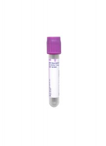
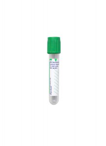
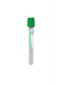
Green BD Vacutainer™ Plus Sodium Heparin Plastic Plasma Tube

Transport Temperature:
Ambient

Specimens must be received by 3 p.m Mondays – Thursdays, and by 12 p.m. on Fridays.

Deliver the specimen to laboratories within 48 hours of collection.

Stability

Ambient: 
48 hours

Refrigerated:
Unacceptable

Frozen:
Unacceptable

Reference Ranges

PHA Stim Index:
≥50.0 SI

Beryllium 1.0 uM D5:
< 3.0 SI

Beryllium 1.0 uM D6:
< 3.0 SI

Beryllium 10 uM D5:
< 3.0 SI

Beryllium 10 uM D6:
< 3.0 SI

Beryllium 100 uM D5:
< 3.0 SI

Beryllium 100 uM D6:
<3.0 SI

Candida albicans:
≥ 2.0 SI

Background Information

Beryllium (Be) is a lightweight metal that can cause acute or chronic diseases that may be asymptomatic. Occupational exposure typically is responsible for this; however, for the most part, it occurs in individuals carrying certain HLA alleles such as HLADPB1 in 83%-97% of individuals with chronic beryllium disease (CBD). CBD is much more common than acute disease nowadays which is typically a chronic non-caseating granulomatous inflammation in individuals who develop beryllium-specific, cell-mediated immunity (typically CD4+ T cells). It is estimated that circa 140,000 workers in the United States are exposed to beryllium every year. Depending on the nature of the exposure and the HLA type, CBD will develop in up to 16% of exposed subjects; thus, CBD remains an important public and occupational health issue. Beryllium-sensitized individuals may remain asymptomatic for life, but approximately 6-8% develop CBD every year.1

Testing for an individual’s sensitivity to beryllium is performed with an in vitro lymphocyte proliferation test (LPT). This is used not only as a screening assay but also as a part of the diagnostic criteria for CBD.

Clinical Indications

Beryllium is a lightweight metal with a high melting point, high strength, and good electrical conductivity. As a result, beryllium has become widely used in a variety of industrial applications, such as thermal coating, nuclear reactors, rocket heat shields, micro-circuits, brakes, X-ray tubes, golf clubs, ceramics, and dental plates. Frequently, it is formulated as an alloy or an oxide.

Correlation between the clinical status of CBD (chronic beryllium disease) and in vitro responses to beryllium in an LPT was developed.2 An elevated blood Be-LPT result may indicate beryllium sensitization, but a definitive diagnosis of CBD requires lung biopsy in the context of compatible signs and symptoms and radiological findings; therefore, as a follow-up step, individuals may undergo pulmonary evaluation, including bronchoscopy, trans-bronchial biopsy, or BAL collection for BAL Be-LPT. This is important in the differential diagnosis of CBD, as sarcoidosis can clinically mimic CBD. Extra-pulmonary CBD may also be occasionally seen such as cutaneous nodules.

Methodology

The blood lymphocyte proliferation test for beryllium sensitization (Be-LPT) is measured by radioactive 3H-thymidine uptake in a cell culture system on two different days after exposure to a range of beryllium sulfate to re-stimulate effector memory CD4+ T cells in peripheral blood. As a control, a common mitogen, phytohemagglutinin (PHA), and Candida antigen are used to ensure acceptable and normal T cell proliferative responses. Emitted beta rays are counted by a beta counter instrument in count per minute (CPM), the stimulation index (SI) is calculated and reported out along with an interpretive comment. The results are reviewed by staff.

Interpretation

The S.I. for PHA should be ≥50.0.

The S.I. for Candida albicans should be ≥2.0 to indicate normal T-cell function.

If all six beryllium indices are less than 3.0, this signifies a normal result, meaning no evidence of beryllium sensitization.

Ratios of greater-than-or-equal-to 3.0 in two or more beryllium indices constitute an abnormal response that is compatible with beryllium hypersensitivity.

References

1. McCanlies EC, Kreiss K, Andrew M, Weston A. 2003. HLA-DBP1 and chronic beryllium disease: a HuGE review. Am. J Epidemiology. 157:388-398.

2. Deodhar SD, Barna BP, Van Ordstrand HS. 1973. A study of the immunologic aspects of chronic berylliosis. Chest. 63:309-313.

3. Barna BP, Culver DA, Yen-Lieberman B, Dweik RA, Thomassen MJ. Clinical application of Beryllium Lymphocyte Proliferation Testing. Clin and Diag Lab Immunol. 2003;10:990-994.

4. U. S. Department of Labor, Occupational Safety and Health Administration. Directorate of Science, Technology and Medicine Office of Science and Technology Assessment, Safety and Health Information Bulletin, September 2, 1999.

June 2019: Changes to UALDOS, TSIG, TRAB

Special Communication

Changes to Aldosterone with Na and K, 24hr Urine (UALDOS), Thyroid Stimulating Immunoglobulin (TSIG), and TSH Receptor Antibody (TRAB) Testing

Changes to Specimen Requirements

Effective May 20, 2019.

Specimen Requirements:

Aldosterone with Na and K, 24-hr Urine (UALDOS)

Volume:
1 mL, 24-hour urine

Minimum Volume:
0.5 mL

Collection Container:
Clean container

Transport Temperature:
Refrigerated

Samples with additives will be rejected.

Changes to Reference Ranges

Effective August 28, 2019.

Interfaced Clients: These updates may require changes to the Test Build.

Reference Ranges:

Thyroid Stimulating Immunoglobulin (TSIG)

Thyroid Stimulating Immunoglobulin:
< 0.55 IU/L

Thyroid Stimulating Immunoglobulin, Qualitative:
Negative

Reference Ranges:

TSH Receptor Antibody (TRAB)

Thyroid Stimulating Immunoglobulin:
< 0.55 IU/L

Thyroid Stimulating Immunoglobulin, Qualitative:
Negative

TSH Binding Inhibition (0 – 99 Years):
< 1.0 U/L

Any additional information will be included in a future Technical Update.  If you have any questions about these changes, please contact Client Services for assistance.

May 2019: Changes to Reference Ranges & Test Builds

Special Communication

May 2019: Changes to Reference Ranges & Test Builds

Changes to Reference Ranges

Effective May 21, 2019.

Reference Ranges:

Cysticercus IgG Ab, Serum (CYSGBL)

≤ 0.8 IV:
Negative
No significant level of cysticercosis IgG antibody detected

0.9 – 1.1 IV:
Equivocal
Questionable presence of cysticercosis IgG antibody detected; repeat testing in 10-14 days may be helpful

≥ 1.2 IV:
Positive
IgG antibodies to cysticercosis detected, which may suggest current or past infection

Effective June 3, 2019.

Reference Ranges:

Liver Fibrosis, FibroTest-ActiTest (LIVFIB)

Apolipoprotein A1

Male:
≥ 115 mg/dL

Female:
≥ 125 mg/dL

Effective June 4, 2019.

Reference Ranges:

Galactose Quant, Plasma (GALAC)

≤ 7 days:
< 5.4 mg/dL

8 – 14 days:
< 3.6 mg/dL

≥ 15 days:
< 2.0 mg/dL

Effective July 2, 2019.

Reference Ranges:

Hemoglobin, A2 (HBA2)

Reference Range
2.0 – 3.1%

Effective July 2, 2019.

Reference Ranges:

Hemoglobin, A2 and F (A2F)

Hemoglobin Fetal:
0.0 – 0.9%

Hemoglobin A2 Percent:
2.0 – 3.1%

Effective July 2, 2019.

Reference Ranges:

Hemoglobin, Fetal (HBF)

Reference Range
0.0 – 0.9%

Effective July 9, 2019.

Reference Ranges:

Vancomycin (VANCRA)

0 – 99 Years:
10.0 – 20.0 µg/mL

Test Build Changes

These updates may require changes to the Test Build for interfaced clients.

Spinal Muscular Atrophy Carrier Screening and Diagnostic (SMAGEN)

Effective May 30, 2019.

Paraneoplastic Autoantibody Evaluation, CSF (PARCSF)

Effective June 11, 2019.

Discontinued Tests

Effective May 21, 2019.

Degradation Products (FDP)

Due to the lack of availability of the appropriate tubes, this test has been discontinued.

We suggest ordering D-Dimer (DDMER).

Any additional information will be included in a future Technical Update.  If you have any questions about these changes, please contact Client Services for assistance.

May 2019: Upcoming Test Changes, Updates, and Discontinuations

Special Communication

May 2019: Upcoming Test Changes, Updates, and Discontinuations

Changes to Reference Ranges

Effective April 24, 2019.

Trypsinogen (TRYPSI)

0 – 17 Years:
Not established

18 Years & Older:
180.5 – 885.3 ng/mL

Changes to Specimen Requirements

Effective May 20, 2019.

Drug Detection Panel, TOF-MS, Umbilical Cord Tissue (DRGTOF)

Volume:
8 inches of umbilical cord (approximately the width of a sheet of paper)

Minimum Volume:
6 inches

Collection Instructions:
Drain and discard any blood. Rinse the exterior of the cord segment with normal saline or water. Pat the cord dry and transport the specimen in a routine urine collection cup or use a Security Kit for Meconium/Umbilical Drug Detection.

Transport Temperature:
Refrigerated

Reference Range:
Gabapentin: Cut-off 10 ng/g

Buprenorphine-G will be removed; all other reference ranges will remain the same.

Drug Detection Panel, Umbilical Cord Tissue, with Marijuana Metabolite (DTOFMP)

Volume:
8 inches of umbilical cord (approximately the width of a sheet of paper)

Minimum Volume:
8 inches

Collection Instructions:
Drain and discard any blood. Rinse the exterior of the cord segment with normal saline or water. Pat the cord dry and transport the specimen in a routine urine collection cup or use a Security Kit for Meconium/Umbilical Drug Detection.

Transport Temperature:
Refrigerated

Reference Range:
Gabapentin: Cut-off 10 ng/g

Buprenorphine-G will be removed; all other reference ranges will remain the same.

Marijuana Metabolite, Umbilical Cord Tissue, Qualitative (DRGTHC)

Volume:
8 inches of umbilical cord (approximately the width of a sheet of paper)

Minimum Volume:
6 inches

Collection Instructions:
Drain and discard any blood. Rinse the exterior of the cord segment with normal saline or water. Pat the cord dry and transport the specimen in a routine urine collection cup or use a Security Kit for Meconium/Umbilical Drug Detection.

Transport Temperature:
Refrigerated

Test Build Changes

These updates may require changes to the Test Build for interfaced clients.

Effective May 20, 2019.

Disopyramide (Norpace) (DISOP)

Drug Detection Panel, TOF-MS, Umbilical Cord Tissue (DRGTOF)

Drug Detection Panel, Umbilical Cord Tissue, with Marijuana Metabolite (DTOFMP)

Ethosuximide (ETHOS)

Discontinued Tests

Effective May 20, 2019.

5-Fluorouracil Toxicity, Chemo Response, 5 Mut (5FLUO)
We suggest ordering Dihydropyrimidine Dehydrogenase (DPYD), 3 Variants (5FUDPD).

Cytochrome P450 2D6 (CYP2D6) Geno (2D6GEN)
We suggest ordering the new test, CYP2D6 (Cytochrome P450 2D6) (2D6GTP).

Cyto P450 2C19 – 9 Variants (2C19PL)
We suggest ordering the new test, CYP2C19 (Cytochrome P450 2C19) (2C19CY).

Warfarin Sensitivity Genotyping (WARSEN)
We suggest ordering new test Warfarin Sensitivity (CYP2C8, CYP2C9, CYP4F2, VKORC1) Genotyping (WRFSEN).

Fats, Urine (UFAT)

Changes to Specimen Requirements

Effective May 29, 2019.

Complement Deficiency Assay (COMPD)

Volume:
1 mL, serum

Minimum Volume:
0.25 mL

Collection Container:
Red (Serum) No Additive Tube

Collection Instructions:
Allow sample to clot, then separate serum from cells as soon as possible. Centrifuge, then remove serum and freeze at minus 20°C or colder (up to 7 days post-draw). Minus 70 °C or colder is preferred and is acceptable up to 30 days post-draw.

Transport Temperature:
Frozen

Tryptase (TRYPT)

Reference Range:
< 8.4 µg/L

Test Build Changes

Effective June 24, 2019.

These updates may require changes to the Test Build for interfaced clients.

HIV-1 Integrase Genotype (HIVIGT)

Any additional information will be included in a future Technical Update.  If you have any questions about these changes, please contact Client Services for assistance.

March 2019: Changes to PR1433, GM1BM, OMEGAC

Immediate Test Changes

Changes to 14-3-3 Protein, CSF (PR1433), Ganglioside Antibody Panel (GM1BM), and OmegaCheck (OMEGAC)

14-3-3 Protein, CSF (PR1433)

Effective March 22, 2019.

Effective immediately, 14-3-3 Protein, CSF (PR1433) is discontinued with no recommended replacement.

The performing laboratory is evaluating new reagents; if these are found to be acceptable, this test may be reactivated in the future.

Ganglioside Antibody Panel (GM1BM)

Effective April 18, 2019.

Ganglioside Antibody Panel (GM1BM) will be discontinued on April 18, 2019.

We suggest ordering Ganglioside Antibodies (GANGAB).

OmegaCheck (OMEGA)

Effective April 22, 2019.

Changes to Reference Ranges

OmegaCheck:
> 5.4% by wt

Arachidonic Acid/EPA Ratio:
3.7 – 40.7

Omega-6/3 Ratio:
3.7 – 14.4

EPA:
0.2 – 2.3% by wt

DPA:
0.8 – 1.8% by wt

DHA:
1.4 – 5.1% by wt

Arachidonic Acid:
8.6 – 15.6% by wt

Linoleic Acid:
18.6 – 29.5% by wt

Any additional information will be included in a future Technical Update.  If you have any questions about these changes, please contact Client Services for assistance.

Herpes Simplex Virus / Varicella-Zoster Virus Detection from Lesions

Technical Brief

Herpes Simplex Virus / Varicella-Zoster Virus Detection from Lesions


Test Name

HSV 1 & 2 / VZV Amplification – Herpes Simplex Virus and Varicella-Zoster Virus, Molecular Detection (HSVVZV)

CPT Codes

87529 – QTY (2)
87798

Methodology

Probe Amplification

Turnaround Time

0 – 3 days

Specimen Requirements

Type:
Swab

Only swabs collected from active cutaneous lesions (vesicles of any skin site) and mucocutaneous lesions are acceptable for testing.

Source:
Genital Lesions (penis; vaginal/cervical)
Skin Lesions
Nares Lesions
Ocular Lesions
Oral Lesions

Specimen Container:
Universal Transport Media (UTM): Regular-Tipped Flocked Swab

CSF Specimens: refer to Herpes Simplex Virus by PCR, CSF (HSPCRC). Transport at ambient temperature up to 30°C for up to 48 hours. Transport on ice pack if warmer temperatures are to be expected.

The following specimen sources are not validated and could be sent out for testing: plasma, serum, amniotic fluid, BAL, and tissue (refer to HSV PCR, Miscellaneous Specimen Types – PCRHSV).

Stability

Ambient: 
48 hours (up to 30°C)

Refrigerated:
7 days (2 – 8°C)

Frozen:
7 days (-20°C)

Background Information

Herpes simplex viruses (HSV) (Types 1 and 2) and Varicella-Zoster virus (VZV) are important causes of vesicular lesions. In many instances, laboratory testing is not needed to prove the presence of the virus; for example, if a patient presents with shingles in a classic dermatomal distribution, then the diagnosis is made based on the clinical findings, and laboratory testing for VZV is not needed. However, in other instances, laboratory testing is essential to demonstrate the presence of the virus (e.g., low-level viral shedding from a mucous membrane-associated lesion). Laboratory studies are also important for differentiation of HSV and VZV when the clinical presentation is not characteristic (e.g. shingles in the genital region).

Clinical Indications

The Symptomatic Patient: This test should be ordered when the cause of vesicular or similar lesion is uncertain.

The Asymptomatic Patient: Asymptomatic vaginal shedding of HSV is a risk factor for the baby during delivery. This test is warranted in pregnant women who are near delivery if there is a concern for the possibility of HSV shedding.

Interpretation

Qualitative test results (i.e. Positive or Negative for HSV1, HSV2, or VZV) will be reported.

Methodology

The Solana HSV 1+2/ VZV Assay is an FDA-approved isothermal amplification assay that detects and differentiates HSV1, HSV2, and VZV. Clinical studies performed at Cleveland Clinic (manuscript in preparation) have demonstrated a 94.7% sensitivity and 100% specificity for the detection of HSV with this assay. The formerly-employed assay (i.e. HSV ELVIS Test System) had a sensitivity and specificity of 71.1% and 93.2%, respectively.

Similarly, the detection of VZV with Solana was superior to direct immunofluorescence (DFA). The sensitivity/ specificity of the Solana assay for the detection of VZV in this study was 95.3%/100%, whereas for DFA these were 71.4%/100%, respectively.

Limitations

This test has limited sensitivity. In rare circumstances, false positives may occur if other intracytoplasmic inclusions are present; however, if the peripheral blood smear is negative, and these diseases are suspected, PCR testing and/or serologic studies are recommended to aid in diagnosis.

References

1. Faron M, Mashock M, Connolly J, Ledeboer N, Buchan B. Evaluation of the Solana HSV 1+2 assay for simultaneous detection of herpes simplex virus 1 and 2, and varicella zoster virus from cutaneous lesions. Session P091; P1894. 27th ECCMID. Vienna, Austria. 22-25 April 2017.

2. Granato PA, Degillo MA, Wilson EM. The unexpected detection of varicella-zoster in genital specimens using the Lyra™ Direct HSV1+2/VZV Assay. J Clin Virol 2016; Nov;84:87-89. doi: 10.1016/j.jcv.2016.10.007. Epub 2016 Oct 11.

3. Huppert JS, Batteiger BE, et al. Use of an Immunochromatographic Assay for Rapid Detection of Trichomonas vaginalis in Vaginal Specimens. Journal of Clinical Microbiology. 2005; 684-687.

4. Solana HSV 1+2/VZV Assay product information and package insert, Quidel, San Diego, CA 92130.

Ehrlichia & Anaplasma Peripheral Blood Smear Review

Technical Brief

Ehrlichia & Anaplasma Peripheral Blood Smear Review


Test Name

Microscopic Examination for Ehrlichia and Anaplasma (EHRLSM)

CPT Codes

87207

Methodology

Giemsa Prepared Thin Smears

Turnaround Time

7 days

Specimen Requirements

Type:
Whole blood

Volume:
5 mL

Minimum Volume:
1 mL

Specimen Container:
Lavender BD Hemogard™ K2EDTA Tube

Transport Temperature:
Ambient

Alternative Specimen

Type:
Whole blood

Volume:
5 mL

Minimum Volume:
1 mL

Specimen Container:
Green BD Hemogard™ Sodium Heparin Tubes

Transport Temperature:
Ambient

Stability

Ambient:
24 hours

Refrigerated:
Unacceptable

Frozen:
Unacceptable

For optimal results, smears must be prepared within several hours of collection.

Background Information

Ehrlichiosis and anaplasmosis are two closely-related tick-borne diseases caused by different and related small obligate intracellular bacteria. Ehrlichiosis is caused by one of several species of bacteria belonging to the genus Ehrlichia, while anaplasmosis is caused by the bacterium Anaplasma phagocytophilum. Both diseases are concentrated east of the Rocky Mountains, with ehrlichiosis found mainly in mid-Atlantic, southeastern, and south-central states, and anaplasmosis occurring most frequently in the Northeast and upper Midwest areas that are endemic for Lyme disease.

Ehrlichiosis and anaplasmosis are emerging infectious diseases in the United States and other countries and were first recognized as illnesses reportable to the CDC in 1999. According to the National Institute of Allergy and Infectious Disease, 1,009 cases of anaplasmosis, 957 cases of ehrlichiosis, and 132 ehrlichial diseases of undetermined type were reported to the CDC in 2008.

Symptoms often begin one to two weeks following an infected tick bite. Clinical manifestations can be mild or life-threatening, resembling those of Rocky Mountain spotted fever, and are often characterized by sudden high fever, fatigue, chills, muscular aches, and headache.

If left untreated, ehrlichiosis can become a severe, life-threatening disease. Severely-ill patients may have low white blood cell count, anemia, elevated liver enzymes, kidney failure, and respiratory insufficiency; other complications can include seizures and coma. Older patients or those with immune suppression are more likely to require hospitalization. Several deaths have been reported.

Possible complications of anaplasmosis include sepsis and damage to the lungs, heart, kidneys, and nerves. Like ehrlichiosis, the disease is noted for being more severe among individuals with compromised immune systems.

Clinical Indications

A patient with feverish symptoms following tick exposure may suspect ehrlichiosis/anaplasmosis. This test is for individuals who are thought to possibly have ehrlichiosis/ anaplasmosis based on symptoms and clinical presentation.

Morulae may be observed in white blood cells with a peripheral blood smear. Although these intracytoplasmic bodies are not frequently seen, their presence is highly-specific if detected by an experienced microscopist.

Interpretation

A positive result indicates the presence of intracytoplasmic morulae. Determining the type of white blood cell that is infected aids in the differentiation between ehrlichiosis and anaplasmosis.

While the specificity of this assay is high, its sensitivity is limited; a negative result does not exclude the possibility of anaplasmosis or ehrlichiosis.

Methodology

Peripheral blood smear (thin film) review following standard Giemsa staining.

Limitations

This test has limited sensitivity. In rare circumstances, false positives may occur if other intracytoplasmic inclusions are present; however, if the peripheral blood smear is negative, and these diseases are suspected, PCR testing and/or serologic studies are recommended to aid in diagnosis.

References

1. National Institute of Allergy and Infectious Disease. Ehrlichiosis and Anaplasmosis. http://www.niaid.nih.gov/topics/ehrlichiosisanaplasmosis/Pages/Default.aspx. Accessed Feb. 1, 2013.

2. Garcia, Lynne S., ed., et. al. Clinical Microbiology Procedures Handbook, 3rd ed., ASM Press, Washington D.C., 2010, Chapters 2.1.17, 3.4.1.17, 11.7.1-11.7.7.

3. Howard, Barbara J., et. al. Clinical and Pathogenic Microbiology, 2nd ed., Mosby Year Book, St. Louis, 1994, pgs. 859-890.

4. Mahon, Connie R., Textbook of Diagnostic Microbiology, 3rd ed., Saunders Elsevier, St. Louis, Mo., 2007, pgs. 1068-1069.

5. Versalovic, James, ed., et al. Manual of Clinical Microbiology, Vol. 1, 10th edition, ASM Press, Washington D.C., 2011, pgs. 240, 1013-1026.

CEBPA Mutation Analysis

Technical Brief

CEBPA Mutation Analysis


Test Name

CEBPA Mutation Analysis, Blood (CEBPA)

CEBPA Mutation Analysis, Marrow (CEBPAM)

CPT Codes

81218

Methodology

Next-Generation Sequencing

Turnaround Time

10 days

Specimen Requirements

Type:
Aspirate, bone marrow

Volume:
2 μg

Specimen Container:
Lavender BD Hemogard™ K2EDTA Tube

Type:
Blood, whole

Volume:
4 mL

Minimum Volume:
2 mL

Stability

Ambient:
48 hours

Refrigerated:
7 days

Frozen:
Unacceptable

Reference Range

CEBPA mutations are not detected.

Background Information

Mutations in the CEBPA gene are identified in 15-18% of acute myeloid leukemia (AML) with normal cytogenetics, and acute myeloid leukemia with mutated CEBPA represents a provisional diagnostic entity in the 2008 WHO classification.[1]

Acute myeloid leukemia with mutated CEBPA displays distinct clinicopathologic features including a favorable clinical course, and the identification of CEBPA mutations may assist in treatment selection.[2-6] CEBPA mutation analysis is recommended for cases of acute myeloid leukemia with normal cytogenetics in the current National Comprehensive Cancer Network (NCCN) and European LeukemiaNet guidelines.

Clinical Indications

Cleveland Clinic Laboratories offers CEBPA mutation analysis for classification and prognostic assessment of new acute myeloid leukemias, especially those with normal cytogenetics. Concurrent NPM1 and FLT3 studies are also recommended.

Interpretation

Mutations in CEBPA include single and dual (usually biallelic) mutations. Initial studies reported that the presence of any CEBPA mutation was associated with a favorable clinical course, while more recent studies have suggested that the favorable clinical course and distinctive clinicopathologic features are limited to acute myeloid leukemia with dual CEBPA mutations.[2-6]

All identified mutations are reported.  Cases are classified as wild type (no mutations detected), single mutated, or dual mutated.

Methodology

DNA is extracted from peripheral blood (CEBPA) or bone marrow (CEBPAM). The entire CEBPA coding region is amplified by PCR and analyzed by Sanger sequencing.

Limitations

Sanger sequencing is expected to identify >99% of mutations, provided that mutations represent at least 15-20% of total CEBPA alleles.

This test is not intended for the detection of minimal residual disease.

References

1. Arber DA et al. (2008). Acute myeloid leukaemia with recurrent genetic abnormalities. In: Swerdlow SH, Campo E, Harris NL et al., eds. WHO Classification of Tumours of Haematopoietic and Lymphoid Tissues. 4th ed. Lyon, France: WHO Press. 110-23.

2. Taskesen E, Bullinger L, Corbacioglu A, et al. Prognostic impact, concurrent genetic mutations and gene expression features of AML with CEBPA mutations in a cohort of 1182 cytogenetically normal AML patients: further evidence for CEBPA double mutant AML as a distinctive disease entity. Blood. 2011;117:2469-2475.

3. Green CL, Koo KK, Hills RK, et al. Prognostic significance of CEBPA mutations in a large cohort of younger adult patients with acute myeloid leukemia: impact of double CEBPA mutations and the interaction with FLT3 and NPM1 mutations. J Clin Oncol. 2010;28:2739-47.

4. Dufour A, Schneider F, Metzeler KH, et al. Acute myeloid leukemia with biallelic CEBPA gene mutations and normal karyotype represents a distinct genetic entity associated with a favorable clinical outcome. J Clin Oncol. 2010;28:570-7.

5. Pabst T, Eyholzer M, Fos J, et al. Heterogeneity within AML with CEBPA mutations: only CEBPA double mutations, but not single CEBPA mutations are associated with favorable prognosis. Br J Cancer. 2009;100:1343-6.

6. Wouters BJ, Lowenberg B, Erpelinck-Verschueren CA, et al. Double CEBPA mutations, but not single CEBPA mutations, define a subgroup of acute myeloid leukemia with a distinctive gene expression profile that is uniquely associated with a favorable outcome. Blood. 2009;113:3088-91.

Amyloid Typing by Liquid Chromatography-Tandem Mass Spectrometry

Technical Brief

Amyloid Typing by Liquid Chromatography-Tandem Mass Spectrometry


Test Name

Amyloid Typing by Tandem Mass Spectrometry (ATMS R)

CPT Codes

88313
88399 – QTY (10)
88380
83789
88321

Methodology

Immunohistochemistry
Liquid Chromatography Mass Spectrometry (LC/MS)

Turnaround Time

2–3 weeks

Specimen Requirements

Type:
Formalin-fixed, paraffin-embedded tissue

Volume:
1 paraffin block

Transport Temperature:
Ambient

Background Information

Systemic amyloidosis is a rare condition characterized by deposits of amorphous material that interfere with the normal structure and function of tissues. Most often amyloid deposits are identified in the heart, kidney, and vascular walls, but virtually every organ system can be affected. All amyloid deposits are thought to be composed of proteins forming beta-pleated sheets, structures that render them insoluble and rigid.

Clinical Indications

Investigations leading to the diagnosis of systemic amyloidosis can be triggered by a variety of symptoms induced by heart or kidney failure, neuropathy, or coagulation abnormalities. Abnormalities suggestive of cardiac amyloidosis have been described by ultrasonography. Amyloid deposits can be identified during workups for low-grade lymphomas, plasma cell myelomas, familial conditions, or renal failure.

There are four major types of systemic amyloidosis: primary amyloidosis, familial amyloidosis, secondary amyloidosis, and senile amyloidosis. The most common form of amyloidosis, primary amyloidosis, is the consequence of deposits of immunoglobulin light chains. Primary amyloidosis is usually diagnosed in patients with plasma cell neoplasms, from monoclonal gammopathy to plasma cell myeloma. Familial amyloidosis is a consequence of inherited mutations, most common in the transthyretin gene. Transthyretin, this time in its wild-type form, is the main component of amyloid in senile amyloidosis, a condition most often diagnosed in the heart. With the decrease in the incidence of chronic inflammatory diseases, mainly infectious, and with better management of autoimmune disorders, the incidence of secondary amyloidosis has markedly decreased and is currently a rare disease. The main amyloidogenic protein in secondary amyloidosis is Amyloid A. Overall, at least 28 proteins have been described to be responsible for the formation of amyloid deposits.

In addition to the proteins that are considered amyloidogenic, amyloid deposits in all types of systemic amyloidosis constantly include other proteins: Serum P component (SAP), Apolipoprotein E (ApoE), Apolipoprotein A-I (ApoA-I), and Apolipoprotein A-IV (ApoA-IV).

Methodology & Interpretation

In histologic sections stained with hematoxylin-eosin, amyloid deposits can be difficult to differentiate from serum or dense collagen. Several stains have been developed to assist in the diagnosis of amyloid, with the Congo Red stain currently considered the gold standard. The microfibrillar nature of the amyloid deposits can be confidently identified by electron microscopy, while the beta-pleated sheet structure of the amyloid can only be demonstrated with x-ray diffraction, a technique exclusively used in research.

While, from a purely morphologic point of view, there are no significant differences between different types of amyloid, the treatment of each type of amyloidosis is different. Immunohistochemical stains show the amyloid deposits to be positive for SAP, but this does not always allow the differentiation of amyloid from serum. In many cases, stains for Transthyretin, Amyloid A, kappa, or lambda immunoglobulin light chains allow further characterization of the components of the amyloid deposits; however, in a significant number of cases, these techniques fail to identify with confidence the amyloidogenic protein. Factors that prevent a confident diagnosis include abnormal protein folding and truncation, as well as the presence of many other endogenous proteins in the amyloid deposits. This results in a significant fraction of cases being inadequately typed and, as a consequence, treated.

Recent studies have shown liquid chromatography-tandem mass spectrometry (LC-MS/MS) to be a reliable method in the identification of amyloidogenic proteins, and is considered by some groups as the current gold standard. This technique was initially used on fresh or frozen tissue, but recently it has been shown that analysis of formalin- fixed, paraffin-embedded tissue (FFPET) can lead to similar results. The analysis usually begins with visual identification of amyloid deposits, followed by their dissection under the microscope (laser microdissection). The specimen is then digested with trypsin, resulting in the generation of peptide fragments. LC-MS/MS is then used to analyze these peptide fragments, resulting in an m/z spectrum. The specific m/z characteristics of the different peaks are compared to those in several databases, leading to the identification of the peptide fragments in the amyloid digested by trypsin. These peptide fragments are quantified and the higher the number of peptide fragments originating from a particular amyloidogenic protein, the higher the degree of confidence that the particular protein is a component of the amyloid. Peptides from the structures of SAP, Transthyretin, ApoE, ApoAI, and ApoA-IV are used as internal controls, indicating the adequate identification of amyloid. When more than one amyloidogenic protein is identified, a comparison of the relative abundance of peptide fragments can indicate which protein is the most abundant in the sample.

This amyloid typing test allows typing of the amyloid deposits with a precision superior to the other available techniques. It incorporates multiple internal controls and allows amyloid typing to be performed in archived specimens. When necessary, in addition to the LC-MS/MS, alternative techniques, such as immunohistochemical stains, can be employed.

Limitations

This test is not a substitute for a surgical pathology consult.

The diagnosis of amyloidosis should not be made by mass spectrometry, as this method has not been developed as a substitute for a detailed morphologic analysis, Congo Red stain, or immunohistochemistry.

Amyloid analysis by tandem mass spectrometry requires that a sufficient amount of amyloid is microdissected. In a few cases, the assay may fail due to the insufficient amyloid available.

In some cases, additional immunohistochemical stains are necessary in order to increase the confidence with which the diagnosis is rendered.

References

1. Sipe JD, Benson MD, Buxbaum JN, et al. Amyloid fibril protein nomenclature: 2012 recommendations from the Nomenclature Committee of the International Society of Amyloidosis. In Amyloid, 2012; 19(4):167-170.

2. Lavatelli F, Vrana JA. Proteomic typing of amyloid deposits in systemic amyloidoses. In Amyloid, 2011; 18(4):177-182.

3. Vrana JA, Gamez JD, Madden BJ, et al. Classification of amyloidosis by laser microdissection and mass spectrometry-based proteomic analysis in clinical biopsy specimens. In Blood, 2009;114(24)4957-4959.

Prostate-Specific Antigen (PSA) – Reference Range Update

Technical Brief

Prostate-Specific Antigen (PSA) – Reference Range Update


Test Name

Prostate Specific Antigen, Diagnostic (PSA)

CPT Codes

84153

Methodology

Electro Chemiluminescence Immunoassay (ECLIA)

Turnaround Time

8 hours

Specimen Requirements

Type:
Plasma

Volume:
1 mL

Minimum Volume:
0.3 mL

Collection Container:
Green BD Hemogard™ Lithium Heparin Tube

Transport Temperature:
Refrigerated

Alternative Specimen Requirements

Type:
Serum

Volume:
1 mL

Minimum Volume:
0.3 mL

Collection Container:
Gold BD Hemogard™ Serum Separation Tubes (SST)™

Transport Temperature:
Refrigerated

Stability

Ambient:
24 hours

Refrigerated:
5 days

Frozen:
24 weeks

Background Information

Prostate-specific antigen (PSA) is a serine protease (~30kDa) secreted almost exclusively by prostate epithelial cells.  PSA was first described in 1979 when it was detected in blood; the concentration of PSA was increased in cases of prostate cancer or other prostatic diseases, such as benign prostatic hyperplasia.[1]

Serum PSA measurement was shown to enhance early detection of prostate cancer, leading to the recognition of its potential as a screening test.[2] Serum PSA alone, or combined with a digital rectal exam (DRE), have been used in clinical trials for early detection of prostate cancer.[3-4] A recent multicenter-study using serum PSA tests with various cutoffs between 2.6 and 4.0 ng/ml showed a 20% reduction in prostate cancer-specific mortality.[4]

Limitations

PSA as a marker is only prostatic tissue-specific rather than cancer-specific. Increased PSA in serum was not only detected in patients with prostate cancer, but also in benign prostatic disease.[1] This phenomenon resulted in a significant false positive rate in PSA-based detection of prostate cancer based on biopsy results from those patients.

Additionally, PSA-based detection of prostate cancer often resulted in over-diagnosis of low-grade cancer that may have never become clinically significant. Several approaches to improve the distinction between cancer and benign conditions have been proposed, including the use of age-adjusted PSA reference ranges, PSA density, PSA velocity, and free-to-total PSA ratio.

Clinical Significance

The traditional cutoff for serum PSA is 4.0 ng/ml. PSA values >4.0 ng/mL were considered abnormal, and these patients were further evaluated with invasive diagnostic approaches, such as prostate biopsy or transrectal ultrasonography of the prostate.[1] However, multiple studies suggest that the risk of prostate cancer in men with PSA levels <4.0 ng/mL is significant (see Table 1). For example, the prostate cancer prevention trial (PCPT) included 2.950 participants who had PSA levels < 4.0 ng/mL and underwent an end-of-study biopsy. Results showed that more than 15% of these men had prostate cancer.[5]

The detection rate of prostate cancer is about 47% in men with serum PSA in the range of 4-10 ng/mL.[5] However, the cancer rate is very similar for men with a PSA range of 2.0-3.0 ng/mL(23%) and that with PSA of 3.0-4.0 ng/ml (26%).

Table 1. Likelihood of Cancer, Based on PSA Level

PSA (ng/mL)

≤ 1.0
1.1 – 2.0
2.1 – 3.0
3.1 – 4.0
4.1 – 10.0
> 10.0

Risk for Prostate Cancer on Biopsy

8.8%
17.0%
23.9%
26.9%
47.0%
58.2%

Data from the prostate cancer prevention trial demonstrated that there is no PSA level below which the risk of having prostate cancer is zero, and suggests that there is no “normal” reference range for PSA. The detection rate of prostate cancer is significantly correlated to the serum PSA levels.

Lowering the PSA cutoff of 4.0 ng/ml to 2.6 ng/ml would increase detection of prostate cancer, while also slightly increasing false positive results.[6-8]

Test Update Information

The new PSA reference range is 0-2.59 ng/ml; the upper limit is cutoff value for further evaluation.

The following comment will be included with each PSA result:

“For an individual patient, the significance of a PSA level should be interpreted in a broad clinical context, including age, race, family history, digital rectal examination, prostate size, results of prior testing (prostate biopsy, free PSA, PCA3), and use of 5-alpha-reductase inhibitors. Considering the high incidence of asymptomatic cancer in the general population that may not pose an ultimate risk to the patient, the decision to recommend urological evaluation or prostate biopsy should be individualized after considering all of these factors.”

A useful tool that incorporates many of these variables for calculating the risk of cancer is available at myprostatecancerrisk.com. This information may assist physicians in deciding whether a prostate biopsy is appropriate.

References

1. Cooner WH, Mosley BR, Rutherford CL, Jr. et al. Prostate cancer detection in a clinical urological practice by ultrasonography, digital rectal examination and prostate specific antigen. J Urol 1990;143:1146-52; discussion 1152-4.

2. Catalona WJ, Smith DS, Ratliff TL et al. Measurement of prostate-specific antigen in serum as a screening test for prostate cancer. N Engl J Med 1991;324:1156-61.

3. Andriole GL, Crawford ED, Grubb RL 3rd, Buys SS, Chia D et al. [PLCO Project Team]. Mortality results from a randomized prostate-cancer screening trial. N Engl J Med. 2009;360(13):1310-9.

4. Schröder FH, Hugosson J, Roobol MJ et al. ERSPC Investigators. Screening and prostate-cancer mortality in a randomized European study. N Engl J Med. 2009;360(13):1320-8.

5. Thompson IM, Pauler DK, Goodman PJ et al. Prevalence of prostate cancer among men with a prostate-specific antigen level < or =4.0 ng per milliliter. N Engl J Med. 2004;350:2239-46.

6. Catalona WJ, Smith DS, Ornstein DK. Prostate cancer detection in men with serum PSA concentrations of 2.6 to 4.0 ng/mL and benign prostate examination. Enhancement of specificity with free PSA measurements. JAMA. 1997;277(18):1452-5.

7. Müntener M, Kunz U, Eichler K et al. Lowering the PSA threshold for prostate biopsy from 4 to 2.5 ng/ml: influence on cancer characteristics and number of men needed to biopsy. Urol Int. 2010;84(2):141-6.

8. Punglia RS, D’Amico AV, Catalona WJ, Roehl KA, Kuntz KM. Effect of verification bias on screening for prostate cancer by measurement of prostate-specific antigen. N Engl J Med. 2003 Jul 24;349(4):335-42.