Streptococcus pneumoniae Antigen Detection in Urine

Technical Brief

Streptococcus pneumoniae Antigen Detection in Urine


Test Name

Streptococcus pneumoniae Antigen, Urine (SPNAG)

CPT Code

87899

Turnaround Time

24 hours

Reference Range

Negative

Specimen Requirements

Volume:
2 mL

Minimum Volume:
0.5 mL

Collection Container:
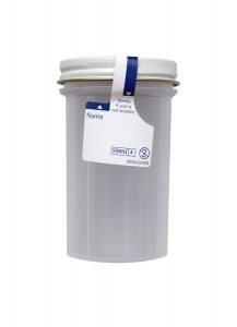
Sterile specimen container

Transport Temperature:
Refrigerated

Stability

Ambient:
24 hours

Refrigerated:
14 days

Frozen:
14 days

Background Information

Streptococcus pneumoniae is the most common organism recovered from patients with community-acquired pneumonia (CAP). Other frequent etiologies of community-acquired pneumonia include Mycoplasma pneumoniae, Haemophilus influenzae, Chlamydophila pneumoniae, and respiratory viruses.

Many different organisms can cause community-acquired pneumonia; risk factors for specific pathogens are available in the consensus guidelines for the management of community-acquired pneumonia, which was jointly developed by the Infectious Diseases Society of America (IDSA) and the American Thoracic Society.[1]

Clinical Indications

Findings suggestive of community-acquired pneumonia (CAP) include productive cough, fever, pleuritic chest pain, hypoxemia, and an imaging study demonstrating a pulmonary infiltrate.

Microbiologic tests to determine the specific cause of community-acquired pneumonia are often negative; however, if the etiology of CAP is determined, empiric therapy can be optimized to decrease morbidity and mortality. De-escalation of therapy can reduce pressure on the emergence of drug resistance and avoid toxic drug effects.

Sputum and blood cultures (prior to initiation of antimicrobial therapy) are recommended for the evaluation of patients with suspected CAP. Recovery of an organism confirms the diagnosis and allows in vitro susceptibility testing to be performed.

Gram stain of sputum is helpful to guide therapy and for correlation with culture results.

Antigen testing is useful when empiric antimicrobial therapy prevents culture confirmation of pneumococcal disease. Urine antigen testing for S. pneumoniae and Legionella pneumophila are recommended for patients with severe CAP.[1]

The S. pneumoniae antigen test has a specificity of >90% and a sensitivity of 50-80% for the diagnosis of pneumococcal pneumonia in adults.[1] However, urinary antigen tests are not recommended for the diagnosis of pneumococcal pneumonia in children because false positive tests are common and attributed to nasopharyngeal colonization with S. pneumoniae.[2, 3]

Methodology

Cleveland Clinic Laboratories uses a qualitative immunochromatographic membrane assay (BinaxNOW® Streptococcus pneumoniae) to detect pneumococcal soluble antigen in human urine. The S. pneumoniae antigen test provides a rapid, simple method for the presumptive diagnosis of pneumococcal pneumonia.

A urine specimen should be collected prior to antimicrobial therapy in a clean, leak-proof container. The specimen may be stored at room temperature if assayed within 24 hours of collection.

Urine is stable for two weeks if refrigerated or frozen. Boric acid may be used as a preservative; however, other preservatives are unacceptable.

Interpretation & Limitations of the Assay

A positive result is presumptive evidence of pneumococcal pneumonia. Correlation of test results with clinical findings is required.

A negative result does not exclude infection by S. pneumoniae since the antigen present in the sample may be below the detection limit of the test.

This test has not been evaluated on patients taking antibiotics for more than one day, or on patients who recently completed a course of antibiotic therapy.[4]

Cross-reactivity with closely-related bacteria in the Streptococcus mitis group may occur.

Streptococcus pneumoniae vaccine may cause false positive results within two days following vaccination, and is not recommended within five days following pneumococcal vaccination.

This test has only been validated for urine samples.

Antigen testing is not recommended for the diagnosis of pneumococcal pneumonia in children.[2, 3]

References

1. Mandell LA, Wunderink RG, Anzueto A, et al. Infectious Diseases Society of America/American Thoracic Society consensus guidelines on the management of community-acquired pneumonia in adults. Clin Infect Dis. 2007 Mar 1; 44 (Suppl 2):S27-72.

2. Dowell SF, Garman RL, Liu G, et al. Evaluation of Binax NOW, an assay for the detection of pneumococcal antigen in urine samples, performed among pediatric patients. Clin Infect Dis. 2001;32:824-825.

3. Bradley JS, Byington CL, Shah SS, et al. The management of community-acquired pneumonia in infants and children older than 3 months of age: clinical practice guidelines by the Pediatric Infectious Diseases Society and the Infectious Diseases Society of America. Clin Infect Dis. 2011;53(t):e25-e76.

4. BinaxNOW® Streptococcus pneumoniae, Package Insert, Alere, Ref. IN710050, Rev. 3, 5/7/10.

March 2019: Changes to Acceptable Clobazam (CLOBAZ) Specimen Types

Immediate Test Changes

Changes to Acceptable Clobazam (CLOBAZ) Specimen Types

Effective March 6, 2019.

Specimen Requirements

Volume:
0.5 mL, serum

Minimum Volume:
0.35 mL, serum

Collection Container:
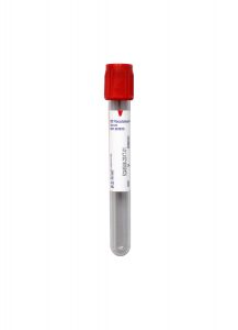
Red BD Hemogard™ Serum Tube (No Additive)

Plasma is no longer an acceptable specimen type.
Serum from a gel tube remains unacceptable.

Specimen Instructions:
Draw specimen immediately before the next scheduled dose. Trough specimens are recommended, as therapeutic ranges are based on trough collections.

Centrifuge the specimen within two hours of collection, transfer the serum into a standard plastic aliquot tube, and transport refrigerated.

Transport Temperature:
Refrigerated

Any additional information will be included in a future Technical Update.  If you have any questions about these changes, please contact Client Services for assistance.

March 2019: Changes to IgG Subclass Reference Ranges

Immediate Test Changes

Changes to IgG Subclass Reference Ranges

Effective March 2, 2019.

IgG Subclass Testing

• IgG Subclass 4 (IGG4)

• IgG Subclasses (IGGSUB)

• IgG Subclasses 1, 2, 3, 4 (IG1234)

Reference Ranges

IgG Subclass 1
10 – 12 Years:  423.0 – 1060.0 mg/dL

IgG Subclass 4:
6 – 8 Years:  0.4 – 99.2 mg/dL

All other reference ranges will remain the same.

Any additional information will be included in a future Technical Update.  If you have any questions about these changes, please contact Client Services for assistance or email Kamran Kadkhoda, PhD, D(ABMLI) with medical questions.

CC-SIGN® NTRK Plus Gene Fusion Next-Generation Sequencing Panel

CC-SIGN® NTRK Gene Fusion Next-Generation Sequencing Panel

This NGS-based laboratory-developed test is designed to detect fusion events in NTRK1, NTRK2, and NTRK3, regardless of fusion partner.

Clinical information obtained from the CC-SIGN® NTRK Gene Fusion NGS Panel can determine if a patient is a candidate for TRK tyrosine kinase inhibitor treatment.

The US Food and Drug Administration (FDA) has approved the use of first-generation TRK tyrosine kinase inhibitors—including larotrectinib (Vitrakvi®) and entrectinib (Rozlytrek®)—to treat patients whose tumors harbor NTRK1, NTRK2, or NTRK3 fusions.

NTRK Fusions are Clinically Actionable

TRK tyrosine kinase inhibitors, such as larotrectinib and entrectinib, are drugs utilized to treat solid tumors with a neurotrophic receptor tyrosine kinase (NTRK) gene fusion without a known acquired resistance mutation. These drugs provide a treatment option to patients with no satisfactory alternative therapies or whose cancer has progressed following treatment, specifically in cases that are either metastatic or where surgical resection is likely to result in severe morbidity.1,2 This treatment targets cancers with NTRK fusions and is not limited by tissue or tumor type.

1. U.S. Food & Drug Administration. FDA approves larotrectinib for solid tumors with NTRK gene fusions. Available at https://www.fda.gov/Drugs/InformationOnDrugs/ApprovedDrugs/ucm626720.htm. [Online]  Updated Dec 17, 2018.
2. U.S. Food & Drug Administration. FDA approves entrectinib for NTRK solid tumors and ROS-1 NSCLC. Available at https://www.fda.gov/drugs/resources-information-approved-drugs/fda-approves-entrectinibntrk-solid-tumors-and-ros-1-nsclc [Online] Updated Aug 16, 2019.

Test Overview

Test Name

NTRK Gene Fusion NGS Panel (NTRK)

CPT Codes

81445, 88381

Turnaround Time

14 days (upon specimen receipt)

Specimen Requirements

Please include the original pathology report with any submitted specimens.

Ten (10) unstained, 4 µM sections of formalin-fixed, paraffin-embedded (FFPE) on charged, unbaked slides

One H&E stained slide with best tumor area circled by a pathologist (minimum of 20% tumor content for best results)

Transport Temperature

Room (ambient) temperature

Specimen Mailing Address

Cleveland Clinic Laboratories
2119 E. 93rd Street, L15
Cleveland, OH 44106

Specimens must be sent via UPS, FedEx, or DHL review our Shipping Information for more details.

Clinical Indications

This test is intended for determining NTRK gene fusion status to identify candidates for larotrectinib treatment.

Limitations

This test does not detect single nucleotide variants; some data show acquired kinase domain resistance mutations that are not interrogated by this test.

Interrogated Genes

NTRK1
NM_002529
Exons 2, 4, 6, 8, 10-14

NTRK2
NM_006180
Exons 5, 7, 9, 11-18

NTRK3
NM_001007156
Exon 15

NTRK3
NM_002530
Exons 4, 7, 10, 12-16

CC-SIGN® Solid Tumor Gene Fusion Next-Generation Sequencing Panel

CC-SIGN® Solid Tumor Gene Fusion Next-Generation Sequencing Panel

This customized, 59-gene Next-Generation Sequencing (NGS)-based laboratory-developed test is intended for use in the diagnosis and management of benign and malignant mesenchymal tumors (sarcomas and their mimics) as well as other solid tumors.

The CC-SIGN® Solid Tumor Gene Fusion NGS Panel is available as part of a comprehensive, diagnostic consultation or as a stand-alone test. This laboratory-developed test interrogates gene targets associated with known translocations in mesenchymal and other solid tumors. Additionally, this panel identifies the corresponding fusion partner and predicted exon breakpoints, which can be of diagnostic and prognostic significance. Fluorescence in situ hybridization-based testing is available for specific fusion questions.

Results are delivered within 14 days of specimen receipt, allowing for a timely, definitive diagnosis in difficult sarcoma and solid tumor cases in a stand-alone fashion or with the support of our expert pathologists.

Test Overview

Test Name

Solid Tumor Gene Fusion NGS Panel (SRCNGS)

CPT Codes

81445
88381

Turnaround Time

14 days (upon specimen receipt)

Specimen Requirements

Formalin-fixed, paraffin-embedded (FFPE) tissue
• Ten (10) unstained, 4 μM sections of FFPE on charged, unbaked slides
• One (1) H&E stained slide with best tumor area* circled by a pathologist

*minimum of 20% tumor content for best results

Transport Temperature

Room (ambient) temperature

Specimen Shipping Address

Cleveland Clinic Laboratories
2119 E. 93rd Street, L15
Cleveland, OH 44106

Specimens must be sent via UPS, FedEx, or DHL review our Shipping Information for more details.

Clinical Indications

This test is intended for the diagnosis of benign or malignant mesenchymal tumors (sarcomas & their mimics) as well as other solid tumors.

Targeted Gene Regions

Genes interrogated, including relevant transcripts and exons, are listed in alphabetical order.

A

Gene, Transcript, Exons

ABL1
NM_004304
2, 4, 6, 10, 16-23, 25, 26

B

Gene, Transcript, Exons

BCOR
NM_001123385
Exons 3-8, 12, 14, 15

BCOR
NM_017745
Exon 8

BRAF
NM_004333
Exons 1-5, 7-16, 18

C

Gene, Transcript, Exons

CAMTA1
NM_015215
Exons 3, 8-10

CCNB3
NM_033031
Exons 2-6

CIC
NM_015125
Exons 12, 17-20

CRTC1
NM_015321
Exons 1-4

CSF1
NM_000757
Exons 5-8

CSF1
NM_172212
Exon 9

E

Gene, Transcript, Exons

EPC1
NM_025209
Exons 9-11

ETV6
NM_001987
Exons 1-7

EWSR1
NM_005243
Exons 4-14

F

Gene, Transcript, Exons

FOS
NM_005252
Exon 4

FOSB
NM_006732
Exons 1, 2

FOXO1
NM_002015
Exons 1-3

FUS
NM_004960
Exons 3-11, 13, 14

G

Gene, Transcript, Exons

GLI1
NM_005269
Exons 4-7

H

Gene, Transcript, Exons

HMGA2
NM_003483
Exons 1-5

J

Gene, Transcript, Exons

JAZF1
NM_175061
Exons 2-4

M

Gene, Transcript, Exons

MAML2
NM_032427
Exons 2, 3

MEAF6
NM_001270875
Exons 4, 5

MKL2
NM_014048
Exons 11-13

MYB
NM_001130173
Exons 7-9, 11-16

N

Gene, Transcript, Exons

NCOA1
NM_147223
Exons 12-15

NCOA2
NM_006540
Exons 11-16

NCOA3
NM_006534
Exons 2, 13-16

NCOA3
NM_181659
Exon 20

NOTCH1
NM_017617
Exons 2, 4, 24-31

NOTCH2
NM_024408
Exons 5-7, 24-29

NOTCH3
NM_000435
Exons 25-30

NR4A3
NM_006981
Exon 2

NR4A3
NM_173200
Exons 3, 4

NTRK1*
NM_002529
Exons 2, 4, 6, 8, 10-14

NTRK2*
NM_006180
Exons 5, 7, 9, 11-18

NTRK3*
NM_001007156
Exon 15

NTRK3*
NM_002530
Exons 4, 7, 10, 12-16

NUTM1
NM_175741
Exons 2-4, 6

*A specimen positive for a fusion in one of these genes makes the patient a candidate for larotrectinib treatment.
Standalone NTRK testing is also available via the CC-SIGN® NTRK Gene Fusion NGS Panel.

P

Gene, Transcript, Exons

PAX3
NM_181459
Exons 6-8

PAX7
NM_002584
Exons 6-8

PDGFB
NM_002608
Exons 2, 3

PDGFD
NM_025208
Exons 5-7

PGR
NM_000926
Exons 1-3

PHF1
NM_024165
Exons 1, 2, 10-12

PLAG1
NM_002655
Exons 1-4

PRDM10
NM_199437
Exons 12, 13

PRKD1
NM_002742
Exons 10-13

R

Gene, Transcript, Exons

RAF1
NM_002880
Exons 4-12

RELA
NM_021975
Exons 3 , 4, 11

RET
NM_020630
Exons 2, 4, 6, 11, 15, 16

RET
NM_020975
Exons 8-14

ROS1
NM_002944
2, 4, 7, 31-38

S

Gene, Transcript, Exons

SRF
NM_003131
Exons 2-4

SS18
NM_001007559
Exons 2-6, 8-11

SS18
NM_005637
Exons 2, 3

STAT6
NM_001178078
Exons 1-7, 15-20

T

Gene, Transcript, Exons

TAF15
NM_139215
Exons 5-7

TCF12
NM_207036
Exons 4-6

TFE3
NM_006521
Exons 2-8

TFEB
NM_007162
Exons 1-4, 9

TFG
NM_006070
Exons 3-7

TRIM11
NM_145214
Exons 2, 3

U

Gene, Transcript, Exons

USP6
NM_004505
Exons 1-3

W

Gene, Transcript, Exons

WWTR1
NM_015472
Exons 3, 4

Y

Gene, Transcript, Exons

YAP1
NM_001130145
Exons 1-9

YWHAE
NM_006761
Exon 5

Solid Tumor Gene Fusion Next-Generation Sequencing Panel

Technical Brief:

Solid Tumor Gene Fusion Next-Generation Sequencing Panel


Test Name

Solid Tumor Gene Fusion Next-Generation Sequencing Panel (SRCNGS) – 59 Genes

Subpanels

Head & Neck Gene Fusion (HDNK) – 30 Genes

NTRK Gene Fusion (NTRK) – 3 Genes

CPT Codes

81445
88381

Methodology

Next-Generation Sequencing

Turnaround Time

14 days

Specimen Requirements

Type:
Formalin-fixed, paraffin-embedded (FFPE) tissue

Volume:
Ten (10) unstained, 4 μM sections of FFPE on charged, unbaked slides

Alternative:
One (1) H&E stained slide with best tumor area circled by a pathologist (minimum of 20% tumor content for best results)

Transport Temperature:
Ambient

Stability

Ambient:
Transport & store at this temperature

Refrigerated:
Unacceptable

Frozen:
Unacceptable

Background Information

Benign and malignant mesenchymal tumors (sarcomas and their mimics) are difficult to diagnose with many benign and malignant entities that differ in their clinical behavior and response to therapy. Many of these tumors harbor gene fusions that are crucial to establishing a definitive diagnosis.

The CC SIGN® Solid Tumor Gene Fusion Next-Generation Sequencing (NGS) Panel is a custom-designed, 59-gene panel, high complexity laboratory-developed test (LDT) designed for targeted sequencing of benign and malignant solid and soft tissue neoplasms. This assay identifies fusion transcripts in targeted regions of RNA from total nucleic acid (TNA) isolated from formalin-fixed, paraffin-embedded (FFPE) tissue specimens.

The test will identify the vast majority of known fusions in benign and malignant mesenchymal tumors, but also has the ability to identify a limitless number of as-of-yet undiscovered gene fusions. This technology only “primes” from one partner of the gene fusion, allowing for the discovery of new gene fusion partners.

Panel Highlights & Available Subpanels

  • Comprehensive detection of gene fusions across 59 targeted genes aids in determining diagnosis, prognosis, and therapeutic options.
  • FFPE tissue removes the need to send fresh or frozen specimens.

Solid Tumor Gene Fusion NGS Panel (SRCNGS) – 59 Genes

  • ALK
  • BCOR
  • BRAF
  • CAMTA1
  • CCNB3
  • CIC
  • CRTC1
  • CSF1
  • EPC1
  • ETV6
  • EWSR1
  • FOS
  • FOSB
  • FOXO1
  • FUS
  • GLI1
  • HMGA2
  • JAZF1
  • MAML2
  • MEAF6
  • MKL2
  • MYB
  • NCOA1
  • NCOA2
  • NCOA3
  • NOTCH1
  • NOTCH2
  • NOTCH3
  • NR4A3
  • NTRK1
  • NTRK2
  • NTRK3
  • NUTM1
  • PAX3
  • PAX7
  • PDGFB
  • PDGFD
  • PGR
  • PHF1
  • PLAG1
  • PRDM10
  • PRKD1
  • RAF1
  • RELA
  • RET
  • ROS1
  • SRF
  • SS18
  • STAT6
  • TAF15
  • TCF12
  • TFE3
  • TFEB
  • TFG
  • TRIM11
  • USP6
  • WWTR1
  • YAP1
  • YWHAE

Subpanel: Head & Neck Gene Fusion (HDNK) – 30 Genes

  • ALK
  • BRAF
  • CAMTA1
  • CRTC1
  • ETV6
  • EWSR1
  • FOS
  • FOSB
  • FOXO1
  • FUS
  • GLI1
  • HMGA2
  • MAML2
  • MKL2
  • MYB
  • NCOA1
  • NR4A3
  • NTRK1
  • NTRK2
  • NTRK3
  • NUTM1
  • PAX3
  • PAX7
  • PLAG1
  • PRKD1
  • RET
  • SS18
  • STAT6
  • TFE3
  • YAP1

Subpanel: NTRK Gene Fusion (NTRK) – 3 Genes

  • NTRK1
  • NTRK2
  • NTRK3

Targeted Gene Regions

Genes interrogated, including relevant transcripts and exons, are listed in alphabetical order.

A

Gene, Transcript, Exons

ABL1
NM_004304
2, 4, 6, 10, 16-23, 25, 26

B

Gene, Transcript, Exons

BCOR
NM_001123385
Exons 3-8, 12, 14, 15

BCOR
NM_017745
Exon 8

BRAF
NM_004333
Exons 1-5, 7-16, 18

C

Gene, Transcript, Exons

CAMTA1
NM_015215
Exons 3, 8-10

CCNB3
NM_033031
Exons 2-6

CIC
NM_015125
Exons 12, 17-20

CRTC1
NM_015321
Exons 1-4

CSF1
NM_000757
Exons 5-8

CSF1
NM_172212
Exon 9

E

Gene, Transcript, Exons

EPC1
NM_025209
Exons 9-11

ETV6
NM_001987
Exons 1-7

EWSR1
NM_005243
Exons 4-14

F

Gene, Transcript, Exons

FOS
NM_005252
Exon 4

FOSB
NM_006732
Exons 1, 2

FOXO1
NM_002015
Exons 1-3

FUS
NM_004960
Exons 3-11, 13, 14

G

Gene, Transcript, Exons

GLI1
NM_005269
Exons 4-7

H

Gene, Transcript, Exons

HMGA2
NM_003483
Exons 1-5

J

Gene, Transcript, Exons

JAZF1
NM_175061
Exons 2-4

M

Gene, Transcript, Exons

MAML2
NM_032427
Exons 2, 3

MEAF6
NM_001270875
Exons 4, 5

MKL2
NM_014048
Exons 11-13

MYB
NM_001130173
Exons 7-9, 11-16

N

Gene, Transcript, Exons

NCOA1
NM_147223
Exons 12-15

NCOA2
NM_006540
Exons 11-16

NCOA3
NM_006534
Exons 2, 13-16

NCOA3
NM_181659
Exon 20

NOTCH1
NM_017617
Exons 2, 4, 24-31

NOTCH2
NM_024408
Exons 5-7, 24-29

NOTCH3
NM_000435
Exons 25-30

NR4A3
NM_006981
Exon 2

NR4A3
NM_173200
Exons 3, 4

NTRK1*
NM_002529
Exons 2, 4, 6, 8, 10-14

NTRK2*
NM_006180
Exons 5, 7, 9, 11-18

NTRK3*
NM_001007156
Exon 15

NTRK3*
NM_002530
Exons 4, 7, 10, 12-16

NUTM1
NM_175741
Exons 2-4, 6

*A specimen positive for a fusion in one of these genes makes the patient a candidate for larotrectinib treatment.
Standalone NTRK testing is also available via the CC-SIGN® NTRK Gene Fusion NGS Panel.

P

Gene, Transcript, Exons

PAX3
NM_181459
Exons 6-8

PAX7
NM_002584
Exons 6-8

PDGFB
NM_002608
Exons 2, 3

PDGFD
NM_025208
Exons 5-7

PGR
NM_000926
Exons 1-3

PHF1
NM_024165
Exons 1, 2, 10-12

PLAG1
NM_002655
Exons 1-4

PRDM10
NM_199437
Exons 12, 13

PRKD1
NM_002742
Exons 10-13

R

Gene, Transcript, Exons

RAF1
NM_002880
Exons 4-12

RELA
NM_021975
Exons 3 , 4, 11

RET
NM_020630
Exons 2, 4, 6, 11, 15, 16

RET
NM_020975
Exons 8-14

ROS1
NM_002944
2, 4, 7, 31-38

S

Gene, Transcript, Exons

SRF
NM_003131
Exons 2-4

SS18
NM_001007559
Exons 2-6, 8-11

SS18
NM_005637
Exons 2, 3

STAT6
NM_001178078
Exons 1-7, 15-20

T

Gene, Transcript, Exons

TAF15
NM_139215
Exons 5-7

TCF12
NM_207036
Exons 4-6

TFE3
NM_006521
Exons 2-8

TFEB
NM_007162
Exons 1-4, 9

TFG
NM_006070
Exons 3-7

TRIM11
NM_145214
Exons 2, 3

U

Gene, Transcript, Exons

USP6
NM_004505
Exons 1-3

W

Gene, Transcript, Exons

WWTR1
NM_015472
Exons 3,

Y

Gene, Transcript, Exons

YAP1
NM_001130145
Exons 1-9

YWHAE
NM_006761
Exon 5

Clinical Indications

This test is intended for the diagnosis of benign or malignant mesenchymal tumors (sarcomas and their benign mimics) as well as other solid tumors.

Interpretation

The results of this test are to be interpreted in the context of the histological, immunohistochemical, and clinical features of the neoplasm.

Methodology

This test relies on Anchored Multiplex PCR (AMP™) technology to generate scalable, target-enriched libraries for NGS from formalin-fixed, paraffin-embedded tissue sections.

In AMP, unidirectional gene-specific primers (GSPs) are used to enrich libraries for known and unknown mutations. Adapters that contain both molecular barcodes and sample indices enable quantitative multiplex data analysis, read de-duplication, and accurate variant calling. Libraries are sequenced on the Illumina MiSeq instrument, which employs “sequencing by synthesis;” a fluorescence, image-based, reversible-terminator technology to sequence targeted regions of the 59 genes included in the panel.

Sequencing data are analyzed for fusion variant detection using Archer® Analysis bioinformatics tools. Specimen quality control is monitored and recorded by in-house developed software (scripts). Raw sequencing data are de-multiplexed based on a unique index sequence using the Illumina bcl2fastq program. The fastq.gz files are de-duplicated according to the unique molecular barcode present and aligned to the human reference genome hg19. Part of the fusion calling and annotation is performed utilizing the Archer® Quiver™ Fusion Database.

Limitations

This test does not detect missense mutations, insertions, deletions, or copy number changes, and does not distinguish between variants that are inherited versus acquired.

References

1. Archer Dx, FusionPlex Anchored MultiPlex PCR (AMP) technology http://archerdx.com/fusionplex/ [Accessed: July 2018]

2. MiSeq System user Guide, Publication Number 15027617 Rev.0. Illumina, San Diego, CA. 9/2014.

3. NCCN Clinical Practice Guidelines in Oncology (NCCN Guidelines®), Soft Tissue Sarcoma, version 1.2019

4. Taylor BS, Barretina J, Maki RG, Antonescu CR, Singer S, Ladanyi M. Advances in sarcoma genomics and new therapeutic targets. Nat. Rev. Cancer. Jul 14 11(8), 541-57 (2011).

December 2018: Changes to RETAB, IA2AB, and PNHPNL Testing

Special Communication

Changes to RETAB, IA2AB, and PNHPNL Testing

Reticulin IgA and IgG Antibodies (RETAB)

Effective December 31, 2018.

Specimen Requirements

Volume:
0.8 mL, serum

Minimum Volume:
0.4 mL, serum

Collection Container:
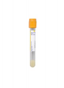
Gold BD Hemogard™ Serum Separation Tube (SST)™

Alternate Container:
Red BD Hemogard™ Serum Tube

Specimen Instructions:
Centrifuge the specimen, then aliquot serum into a standard, non-sterile aliquot tube.

Transport Temperature:
Refrigerated


Insulinoma-Associated Antibody 2 (IA2AB)

Effective January 11, 2019.

Specimen Requirements

Volume:
0.5 mL, serum

Minimum Volume:
0.1 mL, serum

Collection Container:
Gold BD Hemogard™ Serum Separation Tube (SST)™

Transport Temperature:
Frozen

Stability

Ambient:
8 hours

Refrigerated:
48 hours

Frozen:
14 days

Other Information

Methodology:
Enzyme-Linked Immunosorbent Assay (ELISA)

Reference Range:
< 7.5 U/mL

Paroxysmal Nocturnal Hemoglobinuria (PNH) Panel by FCM (PNHPNL)

Effective January 15, 2019.

Stability

Ambient:
48 hours

Refrigerated:
Unacceptable

Frozen:
Unacceptable

Any additional information will be included in a future Technical Update.  If you have any questions about these changes, please contact Client Services for assistance.

Pathology Insights – Pleural Fluid: Cyto-Histo Correlation by Sanjay Mukhopadhyay, MD and Charles Sturgis, MD

Pathology Insights Video Series

Pleural Fluid: Cyto-Histo Correlation

Presented by Sanjay Mukhopadhyay, MD and Charles Sturgis, MD

A cytopathologist (Charles Sturgis, MD) and a thoracic pathologist (Sanjay Mukhopadhyay, MD) discuss their diagnostic approach on a case involving pleural fluid cytology and a concurrent pleural biopsy.

As part of our educational mission for our clients and communities, Cleveland Clinic Laboratories presents the Pathology Insights video series.
These short videos break down information about interesting pathology cases to better inform doctors, laboratory staff, patients, or anyone interested in the field of pathology. Each episode features important cases, methods, and practices that are personally presented by our staff pathologists.

November 2018: Changes to Melatonin (MELAT) Testing

Immediate Test Changes

Changes to Melatonin (MELAT) Testing

Effective November 14, 2018.

Specimen Requirements
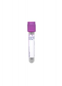
3 mL plasma in a Lavender BD Hemogard™ K2EDTA Tube
Note: Draw two tubes to ensure adequate volume.

• Minimum volume: 1 mL

Specimen Collection
Patient Preparation: Fasting specimen preferred.

Centrifuge the tubes, then transfer plasma into a standard aliquot tube before freezing the specimen.

Transportation
• Transport Temperature: Frozen

Additional Information
Melatonin concentration varies with light and dark cycles: concentration is lower during the day and higher at night.Patients treated with pituitary or steroid hormones influence the concentration.  Hormones should be discontinued for two days prior to collection.

Alternative Specimen Types
• 3 mL serum in a Gold BD Hemogard™ Serum Separation Tube (SST)™

• 3 mL serum in a Red BD Hemogard™ Serum Tube (No Additive)

Any additional information will be included in a future Technical Update.  If you have any questions about these changes, please contact Client Services for assistance.

Hematologic Neoplasm Next Generation Sequencing Panel

Technical Brief:

Hematologic Neoplasm Next-Generation Sequencing Panel


Test Name

Hematologic Neoplasm Next Generation Sequencing Panel – 63 genes
Bone Marrow: HNMNGS   Peripheral Blood: HNPNGS

Subpanels

Myeloid Panel – 50 genes
Bone Marrow: MYNGSM   Peripheral Blood: MYNGSP

Acute Lymphoblastic Leukemia (ALL) Panel – 26 genes
Bone Marrow: ALLBM   Peripheral Blood: ALLPB

Chronic Lymphoproliferative Disorders (LPD) Panel – 7 genes
Bone Marrow: LPMNGS   Peripheral Blood: LPPNGS

Myeloproliferative Neoplasms Panel – 3 genes
Bone Marrow: MPNM   Peripheral Blood: MPNP

CPT Codes

81455

Methodology

Next-Generation Sequencing

Turnaround Time

10 days (upon specimen receipt)

Specimen Requirements

Type:
Bone marrow aspirate

Volume:
2 mL

Type:
Peripheral blood

Volume:
4 mL

Do not freeze specimens.

Stability

Ambient:
48 hours

Refrigerated:
7 days

Frozen:
Unacceptable

Background Information

Recurrent mutations are found in numerous hematologic neoplasms including myelodysplastic syndromes, myeloproliferative neoplasms, acute myeloid leukemia, acute lymphoblastic leukemia, and selected mature lymphoid leukemias.[1-4] The identification of such mutations provides pathologists and clinicians with useful data that may assist in the diagnosis, classification, prognostic evaluation, and therapeutic management of these malignancies. Mutational data in these disorders has been incorporated into the current diagnostic criteria of the World Health Organization Classification of Hematopoietic and Lymphoid Tissues, and into practice guidelines from the National Comprehensive Cancer Network.[5,6]

Cleveland Clinic Laboratories offers a next-generation sequencing panel that analyzes the clinically relevant regions of 62 genes known to be mutated in hematologic neoplasms. This test, performed on peripheral blood or bone marrow aspirate, identifies single nucleotide variants, insertions, and deletions in the targeted genes. Whole-genome copy number analysis may also be obtained by concurrently ordering Cancer Chromosome Microarray + SNP testing.

Smaller subpanels are available for focused disease testing.

Refer to the CC-SIGN® Hematologic Neoplasm Next Generation Sequencing Panel overview for more information.

Panel Highlights & Available Subpanels

Hematologic Neoplasm Next Generation Sequencing Panel – 63 genes

Analyzes the clinically relevant regions of 63 genes known to be mutated in hematologic neoplasms.

  • ABL1
  • ASXL1
  • BCOR
  • BCORL1
  • BRAF
  • CALR
  • CBL
  • CDKN2A
  • CEBPA
  • CSF3R
  • CUX1
  • DDX41
  • DNMT3A
  • EED
  • ETNK1
  • ETV6
  • EZH2
  • FBXW7
  • FLT3
  • GATA1
  • GATA2
  • GNAS
  • IDH1
  • IDH2
  • IKZF1
  • JAK2
  • JAK3
  • KDM6A
  • KIT
  • KMT2A
  • KRAS
  • LUC7L2 (C7orf55)
  • MPL
  • MYD88
  • NF1
  • NOTCH1
  • NPM1
  • NRAS
  • PAX5
  • PHF6
  • PIGA
  • PPM1D
  • PRPF8
  • PTEN
  • PTPN11
  • RAD21
  • RIT1
  • RUNX1
  • SETBP1
  • SF3B1
  • SH2B3
  • SMC1A
  • SMC3
  • SRSF2
  • STAG2
  • STAT3
  • STAT5B
  • SUZ12
  • TET2
  • TP53
  • U2AF1
  • WT1
  • ZRSR2

Subpanel: Myeloid Neoplasm Next Generation Sequencing Panel – 50 genes

Examines 50 genes mutated in myelodysplastic syndromes, myeloproliferative neoplasms, and acute myeloid leukemia.  This panel includes all 34 genes recommended by the Association for Molecular Pathology for analysis of chronic myeloid neoplasms.[7]

  • ABL1
  • ASXL1
  • BCOR
  • BCORL1
  • CALR
  • CBL
  • CEBPA
  • CSF3R
  • CUX1
  • DDX41
  • DNMT3A
  • EED
  • ETNK1
  • ETV6
  • EZH2
  • FLT3
  • GATA1
  • GATA2
  • IDH1
  • IDH2
  • JAK2
  • KIT
  • KMT2A
  • KRAS
  • MPL
  • NF1
  • NPM1
  • NRAS
  • PHF6
  • PIGA
  • PPMID
  • PTEN
  • PTPN11
  • RAD2
  • RUNX1
  • SETBP1
  • SF3B1
  • SH2B3
  • SMC1A
  • SMC3
  • SRSF2
  • STAG2
  • STAT3
  • STAT5B
  • SUZ12
  • TET2
  • TP53
  • U2AF1
  • WT1
  • ZRSR2

Subpanel: Acute Lymphoblastic Leukemia Panel – 26 genes

Includes 26 genes recurrently mutated in lymphoblastic leukemias.

  • ABL1
  • CBL
  • CDKN2A
  • EED
  • ETV6
  • EZH2
  • FBXW7
  • FLT3
  • IKZF1
  • JAK2
  • JAK3
  • KDM6A
  • KMT2A
  • KRAS
  • NOTCH1
  • NRAS
  • PAX5
  • PHF6
  • PTEN
  • RUNX1
  • SH2B3
  • STAT5B
  • SUZ12
  • TET2
  • TP53
  • WT1

Subpanel: Chronic Lymphoproliferative Disorders Panel – 7 genes

Targets seven genes mutated in mature lymphoid leukemias, including chronic lymphocytic leukemia, lymphoplasmacytic leukemia, hairy cell leukemia, and large granular lymphocyte leukemias.

  • BRAF
  • MYD88
  • NOTCH1
  • SF3B1
  • STAT3
  • STST5B
  • TP53

Subpanel: Myeloproliferative Neoplasms Panel – 3 genes

Detects mutations associated with myeloproliferative neoplasms.

  • CALR
  • JAK2
  • MPL

Targeted Gene Regions

Genes interrogated, including relevant transcripts and exons, are listed in alphabetical order.

A

Gene, Transcript, Exons

ABL1
NM_005157.5
Exons 4-6

ASXL1
NM_15338.5
Exons 10-13

B

Gene, Transcript, Exons

BCOR
NM_17745.5
Exons 2-15

BCORL1
NM_021946.4
Exons 1-12

BRAF
NM_004333.4
Exon 15

C

Gene, Transcript, Exons

CALR
NM_004343.3
Exon 9

CBL
NM_005188.3
Exons 8, 9

CDKN2A
NM_000077.4
Exons 1, 2

CDKN2A
NM_058195.3
Exon 1

CEBPA
NM_004364.4
Exon 1

CSF3R
NM_000760.3
Exons 14-17

CUX1
NM_001202543.1
Exons 15-24

CUX1
NM_001913.4
Exons 1-23

D

Gene, Transcript, Exons

DDX41
NM_016222.3
Exons 1-17

DNMT3A
NM_022552.4
Exons 2-23

E

Gene, Transcript, Exons

EED
NM_003797.4
Exons 1-12

ETNK1
NM_018638.4
Exon 3

ETV6
NM_001987.4
Exons 1-8

EZH2
NM_004456.4
Exons 2-20

F

Gene, Transcript, Exons

FBXW7
NM_018315.4
Exons 7-11

FLT3
NM_004119.2
Exons 14-17, 19-20

G

Gene, Transcript, Exons

GATA1
NM_002049.3
Exons 2, 4

GATA2
NM_032638.4
Exons 2-6

GNAS
NM_000516.5
Exons 8-11

I

Gene, Transcript, Exons

IDH1
NM_005896.3
Exon 4

IDH2
NM_002168.3
Exon 4

IKZF1
NM_006060.5
Exons 2, 3, 5-7

J

Gene, Transcript, Exons

JAK2
NM_004972.3
Exons 12-16

JAK3
NM_000215.3
Exons 11-18

K

Gene, Transcript, Exons

KDM6A
NM_021140.3
Exons 1-29

KIT
NM_000222.2
Exons 2, 8-11, 13, 17

KMT2A
NM_005933.3
Exons 1-36

KRAS
NM_004985.4
Exons 2-4

L

Gene, Transcript, Exons

LUC7L2 (C7orf55)
NM_001244585.1
Exons 2-11

M

Gene, Transcript, Exons

MPL
NM_005373.2
Exons 10-11

MYD88
NM_002468.4
Exon 5

N

Gene, Transcript, Exons

NF1
NM_000267.3
Exons 1-57

NF1
NM_001042492.2
Exon 31

NOTCH1
NM_17617.4
Exons 26, 27, 34

NPM1
NM_002520.6
Exons 8-11

NRAS
NM_002524.4
Exons 2-4

P

Gene, Transcript, Exons

PAX5
NM_016734.2
Exons 1-10

PHF6
NM_001015877.1
Exons 2-10

PIGA
NM_002641.3
Exons 2-6

PPM1D
NM_003620.3
Exons 1-6

PRPF8
NM_006445.3
Exons 2-43

PTEN
NM_000314.6
Exons 1-9

PTPN11
NM_002834.3
Exons 3, 4, 12, 13

R

Gene, Transcript, Exons

RAD21
NM_006265.2
Exons 2-14

RIT1
NM_006912.5
Exon 5

RUNX1
NM_001754.4
Exons 2-9

RUNX1
NM_001122607.1
Exon 5

S

Gene, Transcript, Exons

SETBP1
NM_015559.2
Exon 4*

* Exon is only partially analyzed from genomic coordinates chr18:42531679-42532175.

SF3B1
NM_012433.3
Exons 13-16

SH2B3
NM_005475.2
Exon 2

SMC1A
NM_006306.3
Exons 1-25

SMC3
NM_005445.3
Exons 1-29

SRSF2
NM_003016.4
Exons 1, 2

STAG2
NM_00104279.2
Exons 3-35

STAT3
NM_003150.3
Exons 20, 21

STAT5B
NM_012448.3
Exons 16-18

SUZ12
NM_015355.3
Exons 1-16

T

Gene, Transcript, Exons

TET2
NM_001127208.2
Exons 3-11

TP53
NM_000546.5
Exons 2-11

U

Gene, Transcript, Exons

U2AF1
NM_006758.2
Exons 2, 6

W

Gene, Transcript, Exons

WT1
NM_000378.4
Exons 1-9

Z

Gene, Transcript, Exons

ZRSR2
NM_005089.3
Exons 1-11

Clinical Indications

This assay is intended for patients with known or suspected hematologic neoplasms including myelodysplastic syndromes, myeloproliferative neoplasms, acute myeloid leukemia, acute lymphoblastic leukemia, and selected mature lymphoid leukemias.

Interpretation

All variants are classified using Association for Molecular Pathology guidelines for interpretation of somatic variants in cancer.[8]

Detailed interpretations are provided for each variant, and an overall interpretation of the entire mutational profile summarizes the case findings.

Reported variants include those of strong or potential clinical significance as well as variants of unclear clinical significance.

Known benign polymorphisms are not reported.

Methodology

Nucleic acid extracted from the specimen is subjected to nested multiplex PCR-based target enrichment.

Coding and non-coding regions of targeted genes are amplified and sequenced on an Illumina instrument (San Diego, CA) with paired-end, 150×2 cycle reads.

A customized bioinformatic analytical pipeline is used to map reads to the reference human genome (Genomic Build GRCh37/hg19).

Limitations

This test does not detect structural variants or copy number changes and does not distinguish between variants that are inherited versus acquired.

During internal validation, this test delivered an average of >500X coverage and >98% of targeted regions showed over 100X coverage. The test demonstrated 95.2% sensitivity and 99.9% specificity in identifying single nucleotide variants, small insertions and deletions (indels) (≤10bp) of >5% variant allele fraction (VAF). For the identification of large indels (>10bp) at >5% VAF the test demonstrated 87.5% sensitivity and 99.9% specificity.

Due to the limitations of next-generation sequencing technology, some large insertions may not be detected.

References

1. Papaemmanuil E, Gerstung M, Malcovati L, et al. Clinical and biological implications of driver mutations in myelodysplastic syndromes. Blood. 2013 Nov 21; 122(22):3616-27.

2. Bejar R, Stevenson K, Abdel-Wahab O, et al. Clinical effect of point mutations in myelodysplastic syndromes. N Engl J Med. 2011 Jun 30;364(26):2496-506.

3. Döhner H, Weisdorf DJ, Bloomfield CD. Acute Myeloid Leukemia. N Engl J Med. 2015 Sep 17;373(12):1136-52.

4. Nazha A, Zarzour A, Al-Issa K, et al. The complexity of interpreting genomic data in patients with acute myeloid leukemia. Blood Cancer J. 2016 Dec 16;6(12):e510.

5. Arber DA, Orazi A, Hasserjian R, et al. The 2016 revision to the World Health Organization classification of myeloid neoplasms and acute leukemia. Blood. 2016 May 19;127(20):2391-405.

6. “NCCN Guidelines for Treatment of Cancer by Site,” National Comprehensive Cancer Network: https://www.nccn.org/professionals/physician_gls/default.aspx#site.

7. McClure RF, Ewalt MD, Crow J, et al. Clinical Significance of DNA Variants in Chronic Myeloid Neoplasms: A Report of the Association for Molecular Pathology. J Mol Diagn. 2018 Nov;20(6):717-737. DOI: 10.1016/j.jmoldx.2018.07.002. Epub 2018 Aug 20.

8. Li MM, Datto M, Duncavage EJ, et al. Standards and Guidelines for the Interpretation and Reporting of Sequence Variants in Cancer: A Joint Consensus Recommendation of the Association for Molecular Pathology, American Society of Clinical Oncology, and College of American Pathologists. J Mol Diagn. 2017 Jan;19(1):4-23.