Test Name
Trichomonas Prep (TRICHO)
CPT Codes
87808
Methodology
Immunochromatography
Turnaround Time
1 day
Specimen Requirements
Type:
Swab, gential
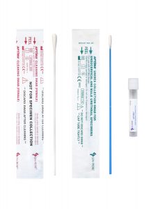
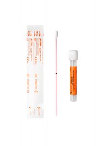
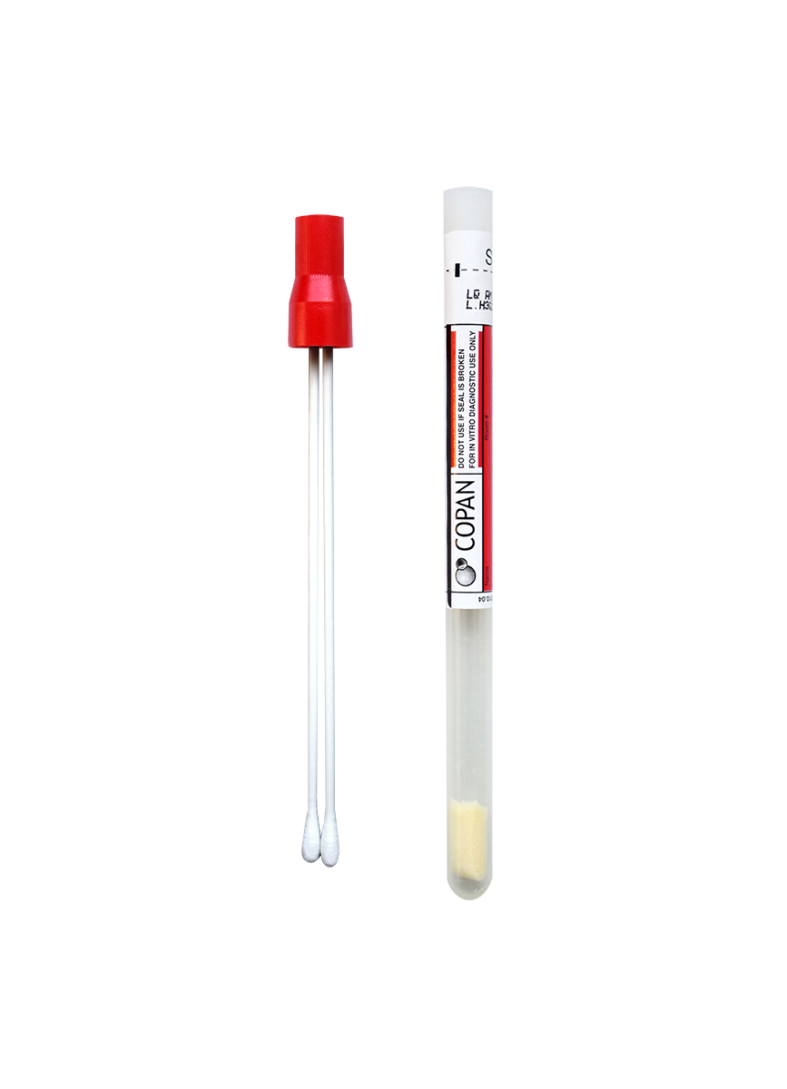
Collection Device:
BD CultureSwab™ Liquid Amies Double Swab
Transport Temperature:
Ambient
Stability
Ambient:
Less than 24 hours
Refrigerated:
36 hours
Frozen:
36 hours
Additional Information
Background Information
Trichomonas is a common cause of vaginitis and the most common nonviral sexually transmitted disease worldwide.1 Studies show that T. vaginalis is an important cause of premature rupture of membranes, premature delivery, pelvic inflammatory disease, urethritis, and chronic prostatitis.
Diagnosis of Trichomonas infection proves challenging in that the traditional wet mount method relies on detecting motility of the parasite, which is often lost during delays in transport or refrigeration. Wet mount preparations are less than optimal when the transport time exceeds six hours.2 Optimal transport time is one hour or less. Alternative methods for diagnosis of Trichomonas infection include EIA methods, in-office physician-performed microscopy, culture, and molecular methods. Molecular diagnostic methods have cost-to-patient charges that are significantly higher than other methods. Wet mount microscopy has a reported sensitivity of 58% versus culture.3
The OSOM® Trichomonas Rapid Test is an FDA-approved, CLIA-waived test that offers a simple, inexpensive option with improved sensitivity for diagnosis of Trichomonas infection as compared to wet prep. The test is intended for qualitative detection of Trichomonas vaginalis antigen from vaginal swabs or from the saline solution prepared when making wet mounts from vaginal swabs.
Clinical Indications
The OSOM® Trichomonas Rapid Test is intended for the qualitative detection of Trichomonas vaginalis antigen from genital swabs. The test is intended for use in patients with symptoms of vaginosis or suspected exposure to the organism.
Interpretation
If Trichomonas is present in the sample, it will form a complex with the primary anti-Trichomonas antibody conjugated to blue-colored particles. The complex will then be bound by a second anti-Trichomonas antibody coated on the nitrocellulose membrane. The appearance of a visible blue test line along with the red control line (internal control) indicates a positive result.
Limitations
The OSOM® Trichomonas Rapid test has not been approved for urine samples; the test has only been validated for qualitative detection of T. vaginalis antigen from vaginal swabs. Molecular methodology is recommended for urine specimens.
A negative result may be obtained if the specimen is inadequate or if the antigen concentration is below the sensitivity of the test.
Samples contaminated with preparations containing iodine or by the immediate prior use of vaginal lubricants are not recommended.
The test does not differentiate between viable and non-viable organisms, nor does it differentiate between acute infection and carrier states.
Staphylococcus aureus in specimens at concentrations higher than 1 X 10⁸ cfu/ml may interfere with the test results in negative samples. These concentrations are higher than would be expected to be present in normal patient samples.
The OSOM® Trichomonas Rapid Test is reported to detect as little as 2500 organisms/ml per manufacturer’s package insert.
Methodology
The OSOM® Trichomonas Rapid Test is an immunochromatographic test that utilizes capillary flow dipstick technology. The test requires solubilization of the Trichomonas proteins from the swab by mixing the sample in the sample buffer. A sample test strip is then added allowing the subsequent migration of the sample along the membrane surface of the dipstick.
References
1. Campbell L, Woods V, et al. Evaluation of the OSOM Trichomonas Rapid Test versus Wet Preparation Examination for Detection of Trichomonas vaginalis Vaginitis in Specimens from Women with a Low Prevalence of Infection. Journal of Clinical Microbiology. 2008; 3467-3469.
2. Huppert JS, Batteiger BE, et al. Use of an Immunochromatographic Assay for rapid Detection of Trichomonas vaginalis in Vaginal Specimens. Journal of Clinical Microbiology. 2005; 684-687.
3. Kingston MA, Bansal D, Carlin EM. Shelf life of Trichomonas vaginalis, International Journal of STD & AIDS. 2003;14:28-29.
4. OSOM® Trichomonas Rapid Test product information and package insert, Sekisui Diagnostics, Framingham, MA, 2011.